Neurocristic hamartoma
Neurocristic hamartoma
Clinical features Neurocristic hamartoma (cutaneous neurocristic hamartoma, pilar neurocristic hamartoma) is an extremely rare developmental, complex hamartomatous lesion of neural crest derivation showing variable differentiation including melanocytic (nevoid, spindled cell, and dendritic components), neurosustentacular (Schwann and perineural cell), and mesenchymal fibrogenic elements.1–11 The skin and superficial soft tissues are affected. Although the majority of these lesions are congenital, acquired variants have rarely been documented.6 Clinically, it generally presents as a localized collection of often folliculocentric, brown, blue or black keratotic macules, papules, and nodules, sometimes with associated alopecia.1,2,6,12 Lesions have been described on the scalp, face, neck, buttock, back, chest wall, and upper extremity.1,2,6,12–14 Some authors have likened the condition to equine melanotic disease.1,3 Occasional reports of melanoma complicating neurocristic hamartoma have been documented.4,5,15 This is often a very late development.15 Such tumors may be relatively indolent and characterized by multiple recurrences over many years or even decades.15 Ultimately, however, metastases develop (particularly affecting the lung) in the majority of cases. Some malignant variants are characterized by very heavy pigmentation and have been described under the rubric pigment synthesizing (animal-type, equine) melanoma.16 Neurocristic hamartoma developing in the background of a giant congenital melanocytic nevus has been reported.17
1300 Melanocytic nevi
seborrheic keratosis-like features.1 Neurocristic hamartoma has also been described in a background of congenital nevus-like features.15
Deep local extension into the skeletal muscle or underlying bone, such as occipital bone with infiltration of bone marrow spaces, can exceptionally be seen in an otherwise ordinary neurocristic hamartoma.14,19
The melanocytic elements express both S100 protein and HMB-45. The Schwann cell nodules express S100 protein and Leu 7 but are HMB-45 negative. They are surrounded by epithelial membrane antigen (EMA)- positive perineural cells and embedded in them are CD34-positive sustentacular cells.2
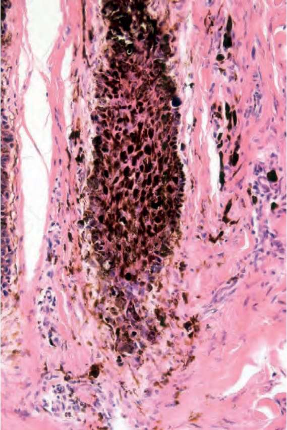
Fig. 25.238 Pilar neurocristic hamartoma: high-power view.
Fig. 25.239 Cellular blue nevus: large dome-shaped lesion with central blue nodule. By courtesy of J.C. Pascual, MD, Alicante, Spain.