Plaquelike blue nevus
Plaquelike blue nevus
Clinical features Plaquelike blue nevus (papular plaque-type blue nevus, eruptive blue nevi, agminate blue nevi) is a rare variant, which may present at birth or develop in children or adults.1–12 It shows a predilection for the trunk but has been described at a variety of sites including the cheek, forearm, breast, foot, scalp, and oral cavity. Clinically, it presents as a usually large (1.3–24 cm) bluish plaque containing multiple darker macules or blue-black papules or nodules. Plaquelike blue nevus has exceptionally been found in association with speckled lentiginous nevus, congenital melanocytic nevus, and atypical Spitz tumor.13
Histologic features Histologically, the plaque-type blue nevus shows variable features. Most commonly, the papules and nodules represent common blue nevi and the intervening macular component shows features reminiscent of a Mongolian blue spot or a nevus of Ota (Figs 25.230 and 25.231). Less often, there are
1297 Dermal melanocytic lesions (dermal melanocytoses)
cellular blue nevus-like features or foci of neurocristic hamartoma.9–12,14,15 Lentiginous hyperplasia affecting the overlying epithelium has also been documented.4
Rare examples of malignant change with features of malignant blue nevus/pigment synthesizing melanoma have been documented.12,16 However, malignant change within plaquelike blue nevus may be difficult to diagnose on morphological grounds alone and further molecular tests including CGH and fluorescence in situ hybridization are indicated to confirm melanoma-associated genomic aberrations.16
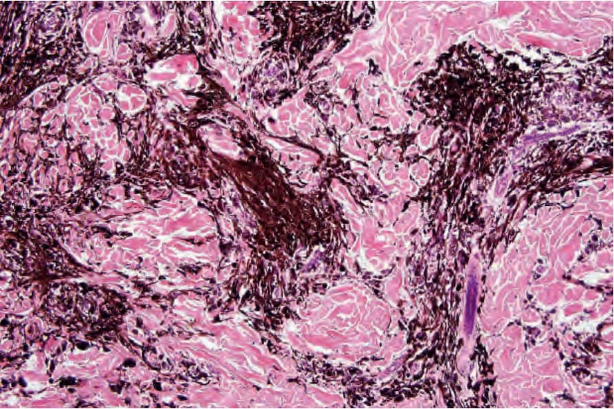
Fig. 25.228 Common blue nevus: medium-power view.
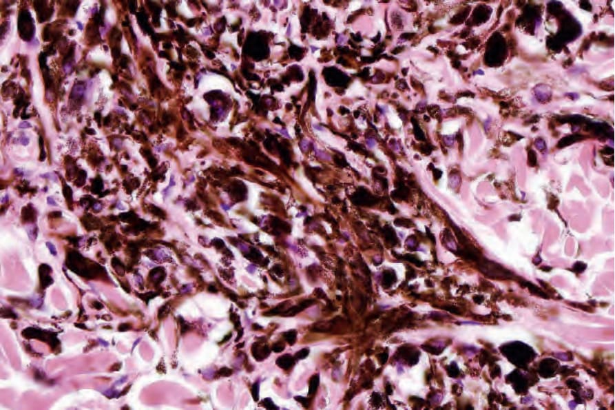
Fig. 25.229 Common blue nevus: high-power view of dendritic cells.
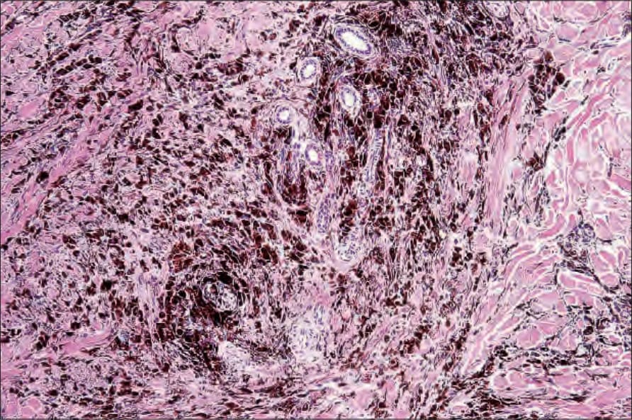
Fig. 25.230 Plaquelike blue nevus: this example shows features of a common blue nevus. By courtesy of K. Busam, MD, Memorial Sloan-Kettering Cancer Center, New York, USA.
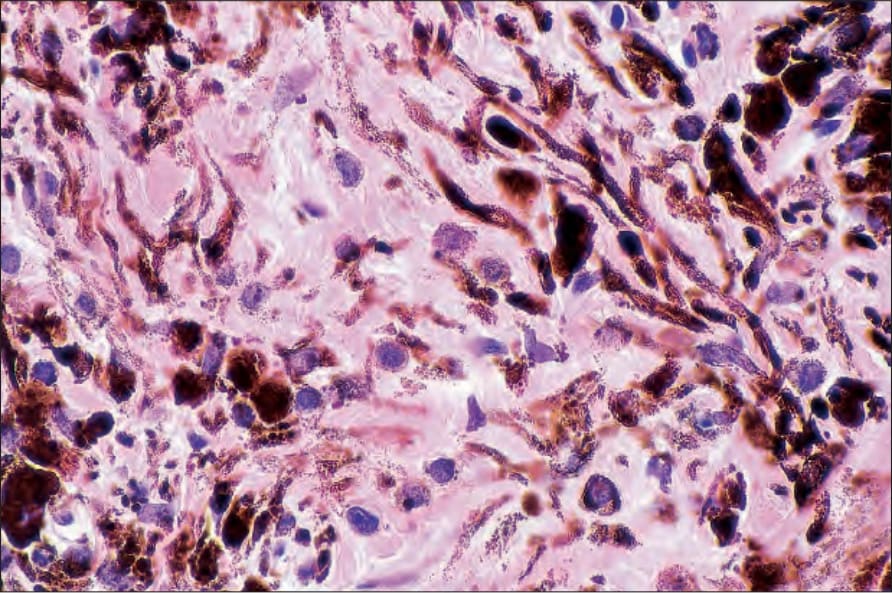
Fig. 25.231 Plaquelike blue nevus: note the heavily pigmented dendritic cells. By courtesy of K. Busam, MD, Memorial Sloan-Kettering Cancer Center, New York, USA.