Giant hairy ‘bathing-trunk’ nevi
Giant hairy ‘bathing-trunk’ nevi
Clinical features Giant hairy ‘bathing-trunk’ nevi are defined as congenital melanocytic nevi measuring 20 cm or more in diameter.1–6 In neonates, melanocytic nevi measuring more than 6 cm on the body and more than 9 cm on the head are considered as giant.7 Giant nevi occur in about 1 : 20 000 neonates.8 They affect the sexes equally and the majority present on the head, neck, and trunk.6 Depending on their exact location, these nevi have been described as ‘bathing-trunk’, ‘vest’, ‘shoulder-sleeve’, ‘stocking’, or ‘glove’ nevi (Figs 25.211–25.213). Satellite lesions are frequently present.4 Giant hairy nevi show variation in color from dark brown to black. Although their color can brighten a few weeks after birth, in most examples, however, it remains unchanged for life.9 Spontaneous regression of giant nevi is exceptional.
Familial occurrence of giant hairy nevi is rare and is possibly linked to polygenic paradominant inheritance.10
A desmoplastic hairless hypopigmented nevus is a variant of giant nevus, characterized by hard consistency, absence of hair, and progressive loss of pigmentation.9,11,12 Cerebriform giant congenital nevus is delineated by cerebriform or gyrate surface pattern of the lesion, and shows predilection for parietal and occipital areas of the scalp.13–15
Giant nevi are, fortunately, extremely rare, since in addition to the disfigurement and psychological trauma they may induce, there is an associated significant risk (3.8–18%) of developing a melanoma.4,6,16 Malignant change usually takes place before puberty and has been reported to be present at birth. There is a striking predilection for axial lesions.6 Giant congenital nevi presenting over the scalp, neck, and posterior midline may have associated leptomeningeal involvement (leptomeningeal or neurocutaneous
1291 Congenital nevi in neonates and young children
neurocristic hamartoma) or even Spitz nevus-like areas.6,36 A coexistent subcutaneous ependymoma has been described.37 Desmoplastic hairless hypopigmented nevus is delineated by prominent dermal fibrosis, progressive disappearance of lesional melanocytes, and hypotrophic or absent hair follicles and sebaceous glands.9
Malignant transformation particularly occurs in the dermal component of the nevus and, in addition to showing a typical melanomatous morphology, tumors may show malignant nerve-sheath tumorlike and anaplastic features.38
Small cell variants and heterologous foci, including rhabdomyosarcomatous and liposarcomatous differentiation, have also been described.6
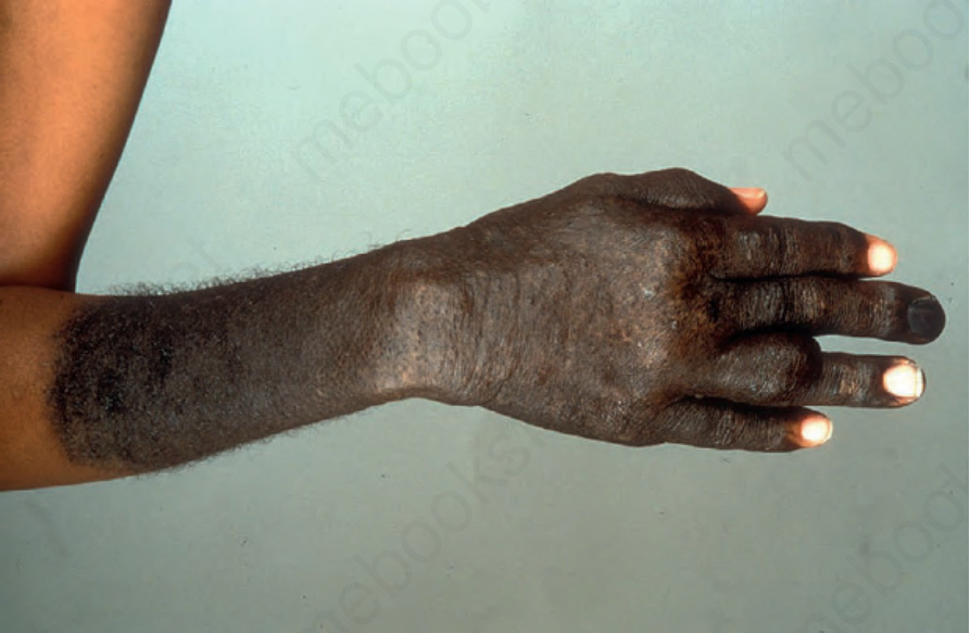
Fig. 25.211 Giant congenital melanocytic nevus: in this example, the nevus has a glove distribution. By courtesy of the Institute of Dermatology, London, UK.
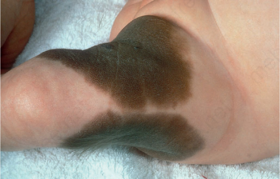
Fig. 25.212 Giant congenital melanocytic nevus: these lesions are very disfiguring and often a source of great concern to the parents. By courtesy of the Institute of Dermatology, London, UK.