Desmoplastic nevus
Desmoplastic nevus
Clinical features Several variants of desmoplastic nevus have been reported – they include a variant with spitzoid cytomorphology (desmoplastic Spitz nevus, hyalinizing spindle and epithelioid nevus), a variant with blue nevus-like morphology, desmoplastic nevus on chronic sun-damaged skin, congenital melanocytic nevus with desmoplasia, and ordinary/common melanocytic nevus with desmoplasia.1–6 Recognition of a desmoplastic nevus is particularly important because it may be confused with a desmoplastic melanoma by the unwary.1–4 Desmoplastic nevus is an entirely benign condition.
Desmoplastic Spitz nevus typically presents as a sometimes scaly, erythematous, or red-brown papulonodule that most often affects the extremities. Although a wide range of age groups may be affected, most patients are in their third decade. Lesions are often of several years’ duration. Desmoplastic melanocytic nevus on chronic sun-damaged skin displays female predominance (70%), most commonly develops on the extremities, and typically presents in the sixth decade of life as a flesh-colored macule or papule.6
Differential diagnosis An irregular growth pattern and lack of uniformity of the nevus population accompanied by significant cytological atypia make a diagnosis of pagetoid Spitz nevus untenable. Fine melanin pigmentation and mitotic activity also favor a diagnosis of melanoma. If the diagnosis is in doubt, a modest
Histologic features There is usually hyperkeratosis and the epidermis often shows acanthosis but sometimes appears normal (Fig. 25.139). The nevus is centered in the papillary dermis but commonly extends into the reticular dermis (Fig. 25.140).2 It consists of a wedge-shaped infiltrate of somewhat pleomorphic cells with abundant eosinophilic cytoplasm containing darkly stained or vesicular nuclei with conspicuous nucleoli (Fig. 25.141).1 Occasionally, residual nests of nevus cells showing spitzoid features are present in the superficial dermis (Fig. 25.142). Intranuclear cytoplasmic pseudoinclusions (invaginations) are often prominent, and sometimes ganglion-like cells are evident (Figs 25.143 and 25.144).2 The nevus characteristically matures
1272 Melanocytic nevi
with depth (Fig. 25.145). Spindled cell, round, and epithelioid forms may all be present (Figs 25.146 and 25.147). Melanin pigment is sometimes seen, but is usually sparse. Mitoses are very infrequent or absent. Occasionally, giant cells with a peripheral ‘wreath-like’ distribution of nuclei are seen.1 The stroma is characteristically extremely dense and often isolates individual nevus cells (Fig. 25.148). Occasionally, there is striking hyalinization – so-called hyalinizing spindle and epithelioid cell nevus.7,8
Junctional activity is sometimes present, but is usually sparse and there is never significant intraepidermal or pagetoid spread. Residual well-formed dermal nests of nevus cells are not often a feature although they may be present in some lesions. Rarely, the nevus may be accompanied by
conspicuous vasculature extending throughout the lesion – the so-called angiomatoid Spitz nevus.9–11 The pathogenesis of the desmoplasia is unknown.
A minority of desmoplastic nevi can feature prominent inflammatory cell aggregates in the mid- or deep dermis composed of lymphocytes and rare plasma cells,6 usually at the periphery of desmoplastic proliferation.6
Differential diagnosis Desmoplastic Spitz nevus is commonly confused with epithelioid fibrous histiocytoma. Junctional activity, melanin pigment, and intranuclear inclusions are not features of epithelioid histiocytoma. In addition, although fibrosis is common, it does not show the extreme degree with often widespread separation of individual tumor cells characteristic of desmoplastic nevus. Desmoplastic nevi are S100 and often HMB-45 positive (Fig. 25.149). Epithelioid
1273 Pigmented spindle cell tumor of Reed
p16 immunohistochemistry can be used as an ancillary method for separating desmoplastic melanoma from desmoplastic nevus. While the majority of desmoplastic melanomas lack or show only weak staining for p16, desmoplastic Spitz nevus consistently displays moderate to strong positivity for this marker.13 Furthermore, p75 immunohistochemistry is usually strongly positive in the majority of desmoplastic melanomas and absent or weak in about 50% of desmoplastic nevi.5
fibrous histiocytoma expresses often positive for ALK-1, and may be focally positive for EMA and smooth muscle actin.
Desmoplastic nevus must also be distinguished from desmoplastic melanoma.12 The latter may show an in situ component (nevertheless, in situ component is absent in about 20–30% of desmoplastic melanomas)6 and is characterized by spindled cells usually distributed in variably sized fascicles and characterized by nuclear basophilia and hyperchromatism. Furthermore, in contrast to desmoplastic nevus, desmoplastic melanoma is usually asymmetrical and frequently extends infiltratively into subcutis.6 Mitotic activity, perineural infiltration, and lymphoid aggregates are also discriminating features and helpful features (see desmoplastic melanoma); however, all of these features can also be found in desmoplastic nevus.
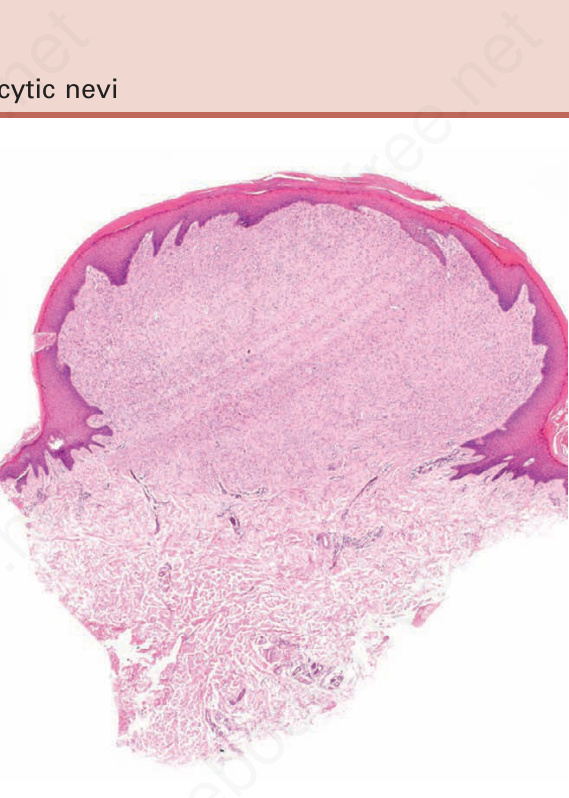
Fig. 25.139 Desmoplastic Spitz nevus: scanning view showing hyperkeratosis, acanthosis, and a well-developed collarette. The nevus predominantly involves an expanded papillary dermis.
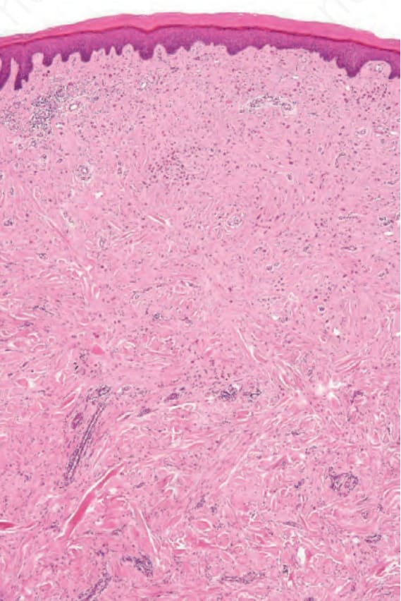
Fig. 25.140 Desmoplastic Spitz nevus: this field shows the full extent of the lesion. There is clear maturation with depth.
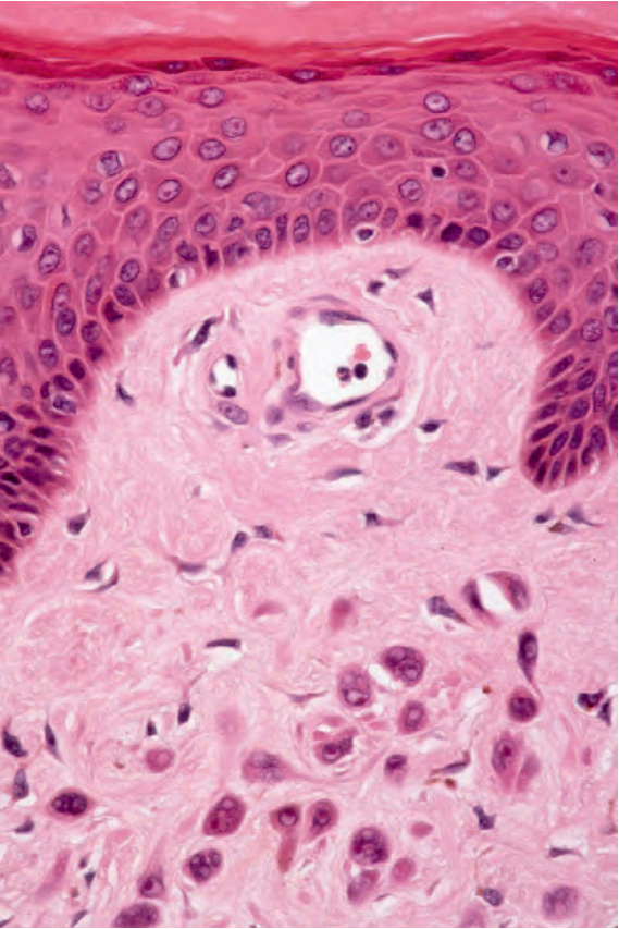
Fig. 25.141 Desmoplastic Spitz nevus: in this example, nevus cells are sparsely distributed in a desmoplastic stroma.
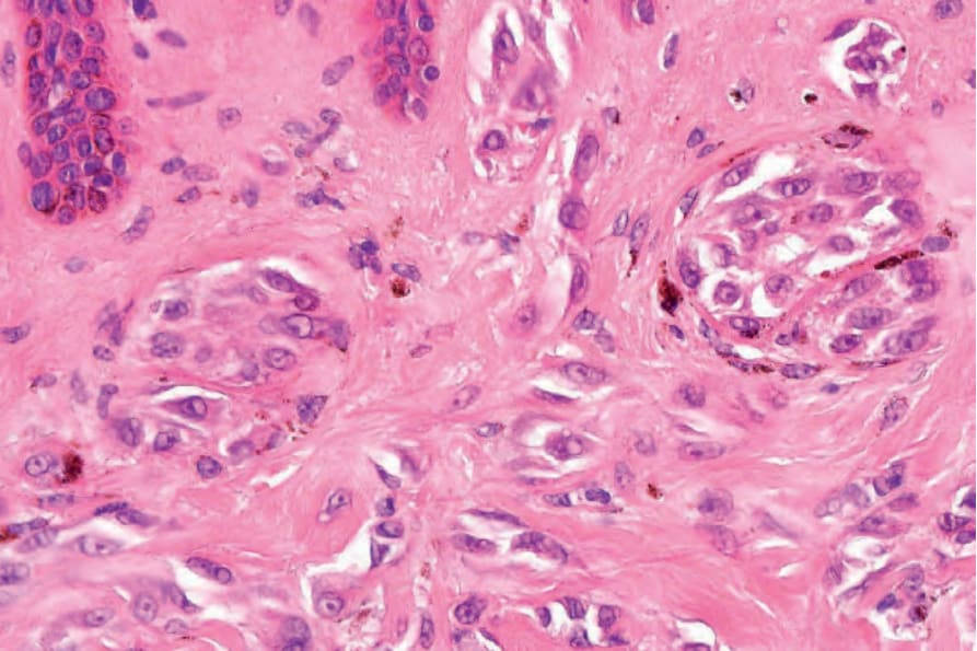
Fig. 25.142 Desmoplastic Spitz nevus: in some examples, the superficial dermal component shows obvious residual, more conventional spitzoid features.
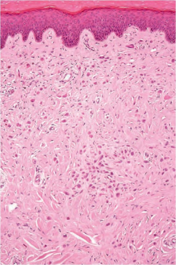
Fig. 25.143 Desmoplastic Spitz nevus: in this more characteristic lesion, the nevus cells are large and have abundant eosinophilic cytoplasm with associated desmoplastic stroma.
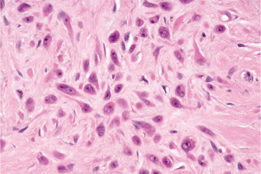
Fig. 25.144 Desmoplastic Spitz nevus: nuclei are vesicular. Cytoplasmic intranuclear pseudoinclusions are prominent.
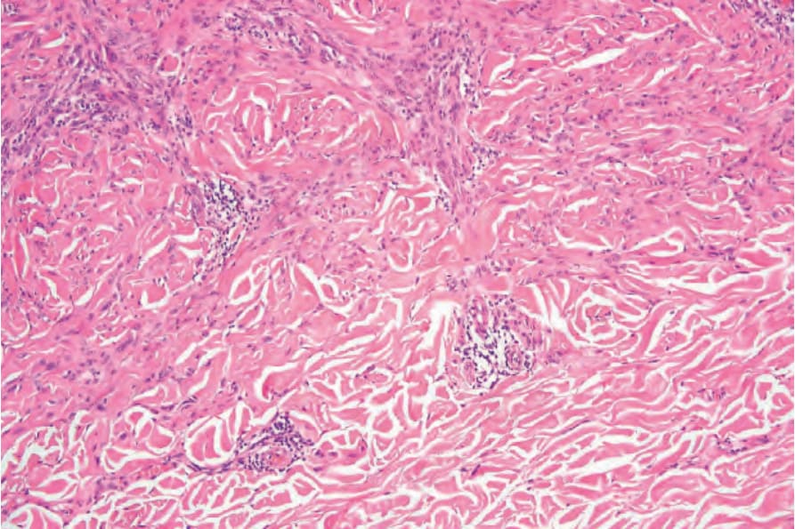
Fig. 25.145 Desmoplastic Spitz nevus: towards the deeper reaches, there is evidence of maturation, as seen by the greatly diminished cell size.
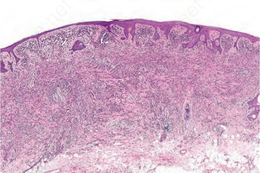
Fig. 25.146 Desmoplastic nevus: low-power view of a spindled cell variant. In this example, the lesion involves the full thickness of the dermis.
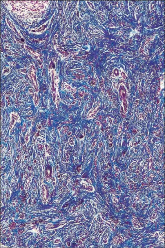
Fig. 25.148 Desmoplastic nevus: the collagenous stroma is highlighted by the Masson trichrome stain.
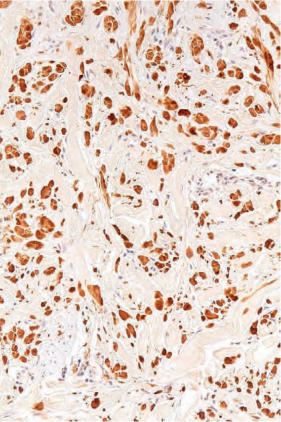
Fig. 25.149 Desmoplastic nevus: S100 protein immunohistochemistry may be helpful in cases where the diagnosis is in doubt.