Nevus spilus
Nevus spilus
Clinical features Nevus spilus (speckled lentiginous nevus) may be congenital but more often it presents in the first year of life. The sexes are involved equally, and there is a predilection for Caucasians. Up to 2% of the population is affected.1 It consists of an aggregate of numerous tiny, pigmented macules and papules arising on a lightly tanned or brown macular background (Figs 25.85 and 25.86).2–4 Nevus spilus ranges in size from less than 1 cm to more than 10 cm and most commonly arises on the trunk and extremities.5 Hypertrichosis has rarely been reported in nevus spilus.6 Lesions are usually solitary. Extensive unilateral (giant) and zosteriform variants have rarely been documented.7–9 Nevus spilus has a tendency to grow along Blaschko lines.10 It may coexist with a plaque blue nevus, bilateral nevus of Ito, or centrofacial lentiginosis and has also been described in association with a nevus sebaceous.11–14 Exceptionally, nevus spilus has also been reported in the oral mucosa.15 Melanoma can, on occasion, develop within a nevus spilus.12,16–25 In such cases, women are particularly affected and the back is most often involved. A speckled lentiginous nevus syndrome has been recognized and consists of a speckled lentiginous nevus in combination with ipsilateral neurological abnormalities such as hyperhidrosis, muscle weakness, and dysesthesia.26 A medial nerve paresis is a recent addition to the syndrome.27
normal, nevoid, or show lentigo simplex-like features.2–4 Cytological atypia may sometimes be encountered.16 Such lesions with worrying features should be carefully monitored for the subsequent development of melanoma.
Nevus spilus has been demonstrated to harbor activating HRAS point mutations.34,35
Histologic features The darkly pigmented speckled areas are characterized by junctional, compound, or dermal nevi. Less often, Spitz nevi and blue nevi also in an agminate pattern may be seen.28–33 The intervening background skin may be
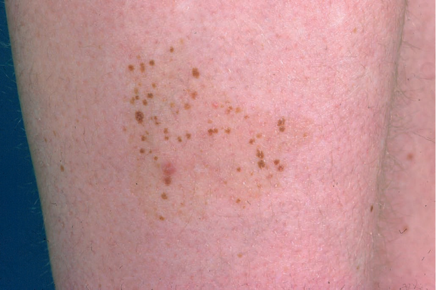
Fig. 25.85 Nevus spilus: heavily pigmented macules are present within a background circumscribed paler lesion. From the collection of the late N.P. Smith, MD, the Institute of Dermatology, London, UK.
Fig. 25.87 Cockarde nevus: note the characteristic targetoid appearance. By courtesy of J.C. Pascual, MD, Alicante, Spain.