Clonal nevus
Clonal nevus
Clinical features Many so-called clonal nevi (inverted type A nevi) are clinically unremarkable. Those that are recognized are characterized by a recent change, usually in color, in an otherwise typical banal or (less commonly) congenital nevus.1,2 Alternatively, they may exhibit darker pigmentation in the background of an otherwise uniformly pigmented nevus.3
On dermoscopy, clonal nevi are characterized by a uniform globular/ cobblestone pattern typically containing an eccentric fairly uniform blue gray blotch.4,5
Histologic features This variant of nevus is characterized by the presence of a usually circumscribed nest or collection of nests in the superficial dermis distinct from the background nevoid population (Figs 25.61–25.64). The melanocytes are typically epithelioid, with often abundant, heavily or finely pigmented cytoplasm and irregular nuclei containing small nucleoli.1,2 Melanophages are usually numerous in the adjacent dermis, but a lymphocytic response is absent. Mitoses are absent or extremely rare. The nests stand out against the background population of type B nevus cells – hence the designation inverted type A nevus.
Histologic features The nevus is characterized by a striking syringocentric distribution. The sweat duct epithelium may be involved but the secretory unit is typically unaffected. The overlying and adjacent epidermis shows features of a lentigo.
Differential diagnosis The vast majority of clonal nevi most likely represent combined or deep penetrating nevi. Melanocytic nevi with a focal atypical epithelioid component (clonal nevus) share similar age, anatomic distribution, and cytological features with the deep penetrating nevus, but lack the deep extension of melanocytes.6
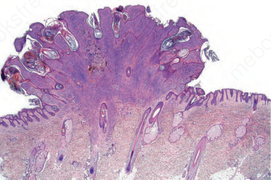
Fig. 25.61 Clonal nevus: this is a banal compound nevus. Note the distinct nodule in the deeper dermis surrounded by pigment-laden melanophages. By courtesy of W. Grayson, MD, National Health Laboratory Service, Johannesburg, South Africa.
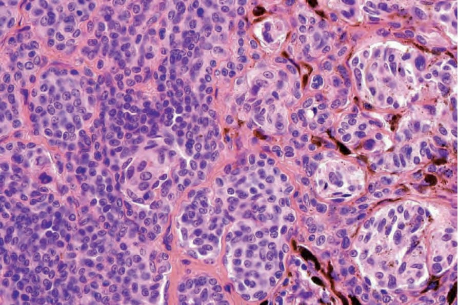
Fig. 25.63 Clonal nevus: high-power view of type A nevus cells with fine pigmentation. By courtesy of W. Grayson, MD, National Health Laboratory Service, Johannesburg, South Africa.