Melanocytic nevus
Melanocytic nevus
melanophages, and a mild perivascular chronic inflammatory cell infiltrate is sometimes present.
In occasional lesions, the reticular dermis contains large numbers of hamartomatous, irregular, enlarged smooth muscle fibers (Fig. 25.21).25–28 Associated dermal fibrosis, sebaceous hyperplasia, neves sebaceous, localized acneiform lesions, solitary plexiform neurofibroma, neurofibromatosis type I, basal cell carcinoma, compound melanocytic nevus, pigmented epithelioid melanocytoma, nevus anemicus, lichen planus, pityriasis versicolor, a psoriasiform dermatitis suggestive of an inflammatory linear verrucous epidermal nevus, and hyperkeratosis with focal diminution of granular cell layer (e.g., ichthyotic changes) have also been documented.3,28–38
Melanocytic nevus (banal nevus) is a benign tumor that usually presents in childhood and adolescence. The sex distribution is equal. An average white individual can expect to develop 15–40 such lesions during life, reaching the maximum number in the third decade before regression to virtual disappearance by the eighth and ninth decades.1–5
In addition to those derived from hair-bearing skin, melanocytic nevi may develop on glabrous skin, beneath fingernails and toenails, within the conjunctivae and uveal tract, and mucosae (Fig. 25.22). They have an ordered and histologically defined natural history, which may, to some extent, be predicted from their clinical appearance. Their existence commences as a focus of melanocytic proliferation (junctional activity) within the lower reaches of the epithelium, the so-called junctional nevus. This progresses to the presence of melanocytes within both the epidermis and the dermis, which is the compound nevus. Further development results in a completely intradermal lesion called the dermal nevus.6
Ultrastructural studies have revealed an increased number and size of compound melanosomes within basal keratinocytes.3 An increase in the number of melanosomes per complex may also be evident.
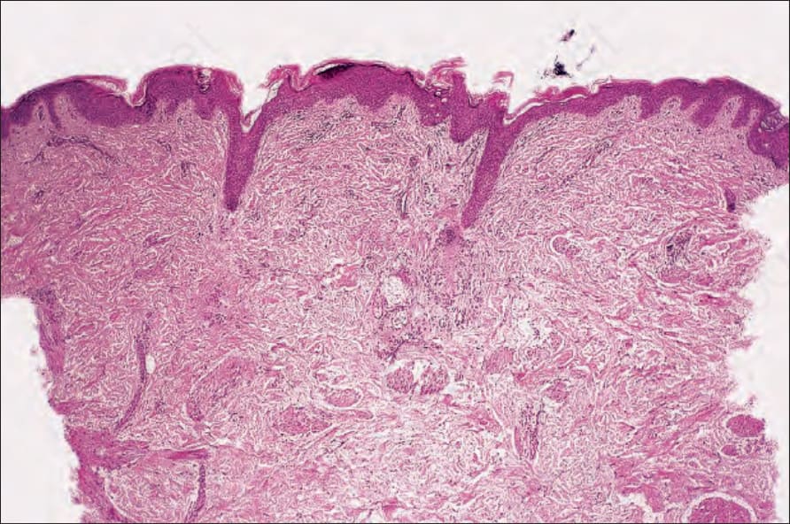
Fig. 25.21 Becker nevus: in this example, hamartomatous aggregates of smooth muscle cells are widely distributed throughout the dermis.
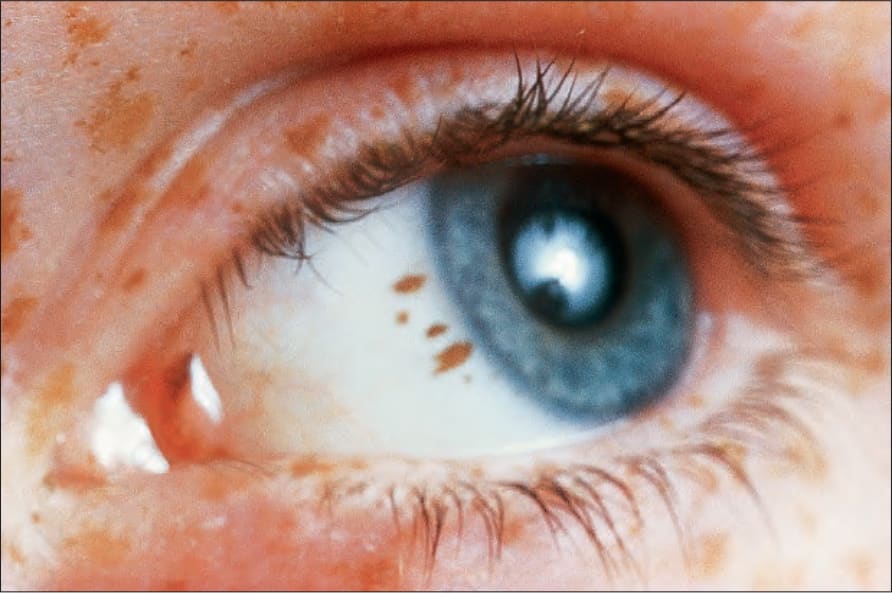
Fig. 25.22 Conjunctival junctional melanocytic nevus: there is uniform pigmentation and a regular border. By courtesy of the late M. Beare, MD, Royal Victoria Hospital, Belfast, N. Ireland.