PUVA and sunbed lentigines
PUVA and sunbed lentigines
Clinical features Long-term psoralen photochemotherapy (PUVA) may be complicated by a variety of cutaneous pigmentary changes including variable hyper- and hypopigmentation, vitiligo-like features, and multiple lentigines.1–5 PUVA lentigines are dose-dependent, irregular, small, brown-black macules that are particularly seen on the shoulders, upper back, and limbs (Fig. 25.13).3,4 They are commonly numerous. Males are more often affected than females, and skin types I and II are particularly at risk. Similar lesions have occasionally been described in patients, including one with systemic lupus erythematosus, following the use of sunbeds for artificial tanning.6–8 Lesions tend to regress after therapy is discontinued.4 Development of PUVA lentigines has been reported at the sites of mycosis fungoides lesions, as well as in normal and vitiliginous skin.9,10 Furthermore, narrow band ultraviolet (UV) B used in patients with early-stage mycosis fungoides induce development of lentigines in both involved and non-involved skin much earlier and at lower cumulative dose than those developing after PUVA treatment. However, lentigines develop less frequently than those presenting after PUVA treatment.11
Pathogenesis and histologic features The presence of T1799 BRAF mutations has been demonstrated in 33% of PUVA lentigines.12
Histologically, PUVA lentigines show a variety of features.1–5,13,14 In some, there is increase in basal cell pigmentation with no increase in melanocyte numbers, reminiscent of an ephelis, whereas others resemble lentigines with pronounced rete ridge elongation and increased numbers of melanocytes (Fig. 25.14). Melanocytic nuclear atypia including enlargement, pleomorphism, and hyperchromasia, multinucleation, and giant melanosomes has been described.13,14 There does not appear to be any link between PUVA lentigines and development of melanoma.5
eyes (skin types I and II) and presents on a background of solar-damaged skin as a usually solitary, irregular, reticulated black macule with a wiry or beaded appearance (reminiscent of an ink spot) (Fig. 25.15).1–3 Although the features may be clinically worrying, the lesion is completely benign. Ink spot lentigo can exceptionally develop in the background of a nevus spilus.4
Epidermal abnormalities, including dyskeratosis and actinic keratosis-like features, may also sometimes be observed.15
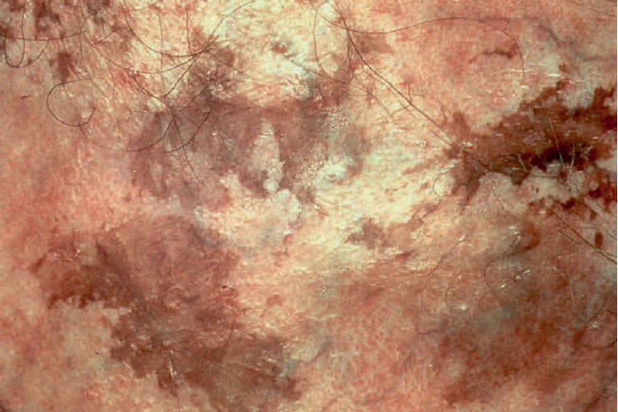
Fig. 25.13 PUVA lentigines: multiple lentigines are evident against a background of variable hypo- and hyperpigmentation. By courtesy of the Institute of Dermatology, London, UK.
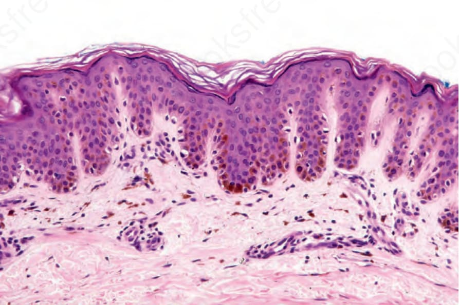
Fig. 25.14 PUVA lentigo: the features are indistinguishable from a lentigo simplex.
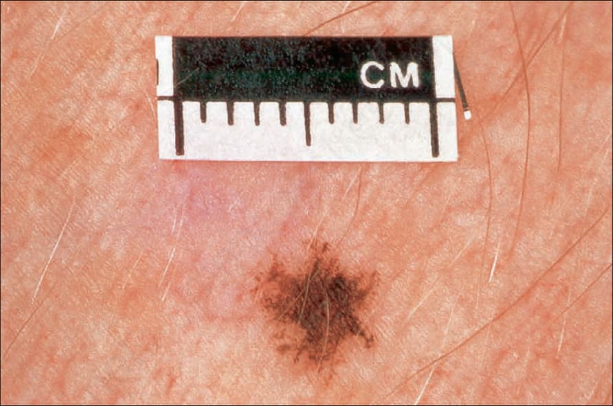
Fig. 25.15 Ink spot lentigo: this is a typical extremely irregular pigmented macule. By courtesy of the Institute of Dermatology, London, UK.