Glomus tumor
Glomus tumor
Clinical features Glomus tumor arises in young adults and is usually located in the distal extremities, with a predilection for the fingers. The single most common site is the subungual region,1 where a female predominance is observed.2
Malignant glomus tumors and glomus tumor of uncertain malignant potential are usually not encountered in a subungual location (see Glomus tumor, Chapter 35). Nevertheless, symplastic glomus tumor has been described in the nail apparatus. This benign variant is characterized by high-grade nuclear atypia in the absence of any other malignant features. The atypia is thought to represent a degenerative phenomenon.14
Differential diagnosis The differential diagnosis encompasses eccrine spiradenoma and hidradenoma. Both are characterized by focal ductal differentiation and positivity for epithelial markers.
1154 Diseases of the nails
Clinical features Subungual exostosis is a benign lesion that occurs exclusively in the nail region. It presents as a slowly growing, flesh-colored or erythematous, tender nodule under the nail plate. Subungual hyperkeratosis, onycholysis, nail deformity, superficial erosion, or ulceration may be observed (Fig. 23.73).
The lesion usually occurs in the second and third decades of life. There is a strong predilection for the toes, especially the great toes. Involvement of the fingers is rare.1 An X-ray scan shows a mineralizing tumor attached to the distal phalanx in the absence of cortical and medullary continuity between the tumor and the underlying bone (Fig. 23.74).2 Complete excision is the recommended treatment.
Pathogenesis and histologic features Subungual exostosis was thought to be a reactive process; however, recent genetic data suggest that these are probably true neoplasms. The tumors contain a t(X;6) translocation.3–6 The lesion is typically composed of a bony stalk lined by osteoblasts and covered by a cartilaginous cap (Figs 23.75 and 23.76). There is a spindle cell proliferation that matures into cartilage. The cartilage, which appears proliferative with increased cellularity and binucleate forms, in turn, matures into trabecular bone. The intertrabecular spaces contain adipocytes and loosely arranged spindle cells.6
1155 Soft tissue and bone tumors
Differential diagnosis Osteochondroma, the most common benign bone tumor, usually arises in the femur, humerus, and tibia. It is extremely unusual in a subungual location. It is a projection from the surface of the bone, with its cortex and spongiosa continuous with the cortex and the spongiosa of the underlying bone. Subungual exostosis is attached to the distal phalanx but shows no such continuity. Osteochondroma is a bony projection capped by hyaline cartilage. It lacks the spindle cell proliferation which matures into cartilage, encountered in subungual exostosis.
Enchondroma and periosteal chondroma are cartilaginous neoplasms that do not form bone.5
Access ExpertConsult.com for the complete list of references
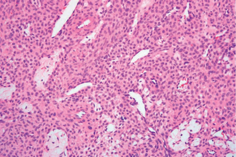
Fig. 23.72 Glomus tumor: medium-power view showing thin-walled dilated vessels surrounded by typical glomus cells.
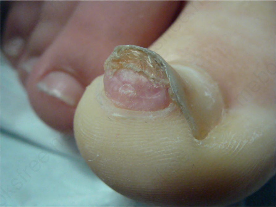
Fig. 23.73 Subungual exostosis: hyperkeratotic tumor lifting up the distal nail plate. Courtesy of B. Richert, MD, PhD, Université Libre de Bruxelles, Belgium.
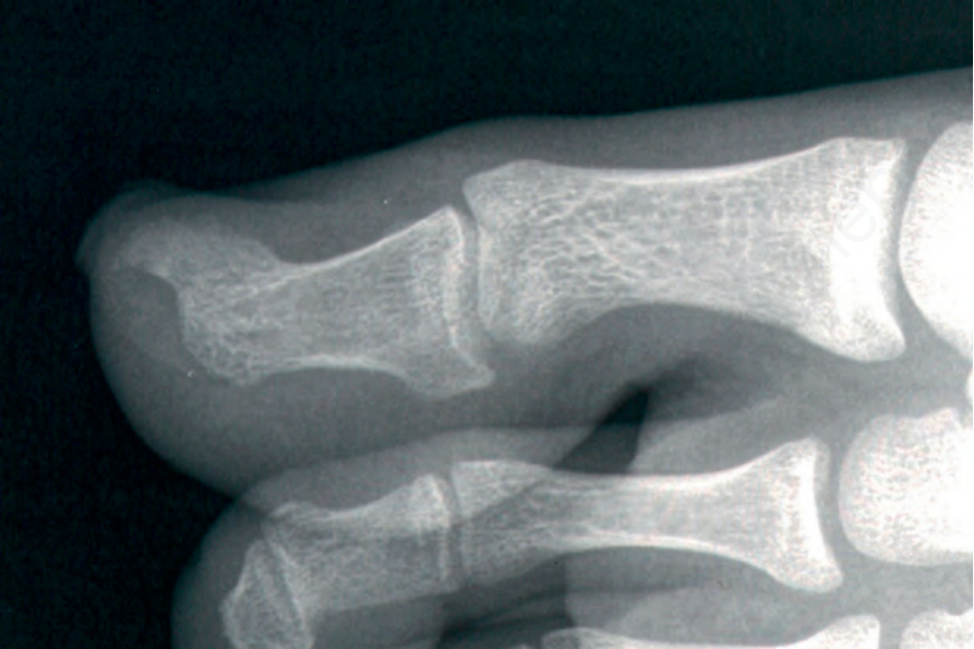
Fig. 23.74 Subungual exostosis: there is an irregular bony tumor deep to the nail plate. Courtesy of B. Richert, MD, PhD, Université Libre de Bruxelles, Belgium.
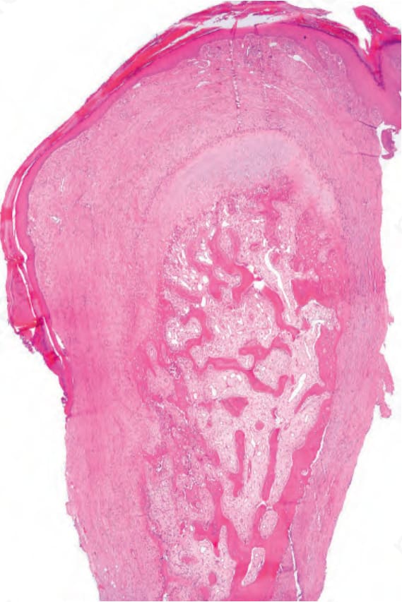
Fig. 23.75 Subungual exostosis: scanning view of an oval bony stalk covered by a cartilaginous cap with overlying thin, hyperkeratotic epidermis. Subungual exostosis
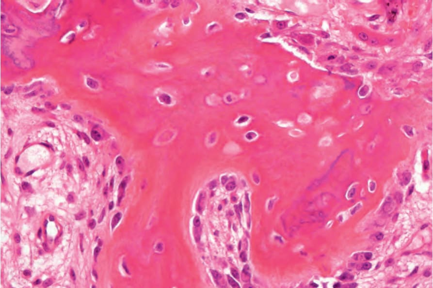
Fig. 23.76 Subungual exostosis: high-power view of the bony stalk.