Myxoid pseudocyst
Myxoid pseudocyst
Clinical features Myxoid pseudocyst (digital mucous cyst) was initially thought to represent focal myxoid degeneration of connective tissue. However, there is
now some evidence to suggest that it results from escape of synovial fluid through a breach in the synovial capsule.1 It is usually located in the PNF and occurs in middle-aged or elderly patients.2 It is about 10 times more frequent on the fingers than on the toes. The lesion starts as an asymptomatic swelling which slowly enlarges up to 2 cm. The cyst contains clear viscous fluid. It characteristically causes a longitudinal depression in the nail plate. Lesions can be solitary or multiple and can be job related.3–5 Subungual location associated with transverse overcurvature of the nail plate has also been reported.6 Recurrence after incomplete excision is not uncommon.
1153 Soft tissue and bone tumors
Histologic features Myxoid pseudocyst is well circumscribed but unencapsulated dermal and devoid of any lining epithelium. It consists of a large mucin-filled space containing spindle-shaped and stellate fibroblasts without atypia. The mucin contains mucopolysaccharides, which stain positively with Alcian blue and colloidal iron.7
Differential diagnosis Ganglion cyst is usually subcutaneous, and occasionally a connection between the cyst and the underlying joint cavity can be demonstrated.7 It consists of myxoid spaces incompletely lined by flattened synovial cells and surrounded by a fibrous wall.8
Superficial angiomyxoma is larger, lobulated, and characterized by more abundant spindled-shaped and stellate cells, frequent small blood vessels, and collections of neutrophils.9
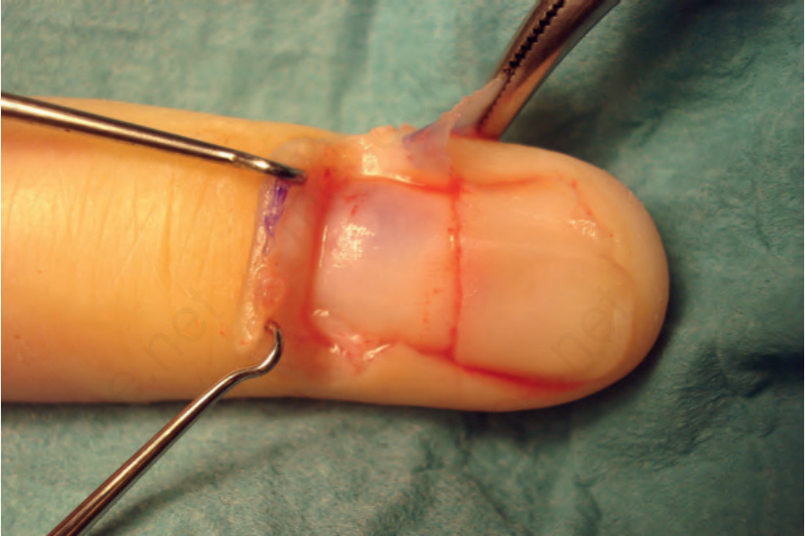
Fig. 23.71 Glomus tumor: there is an ill-defined bluish tumor deep to the proximal nail plate. Courtesy of B. Richert, MD, Université de Liège, Belgium.