Onychopapilloma
Onychopapilloma
Differential diagnosis The diagnosis of onychopapilloma may be difficult when the nail plate had been avulsed and put back in place during the surgical longitudinal excision, with most of the lesion remaining attached to the nail plate.5
Pathological examination of localized (isolated) longitudinal erythronychia may reveal onychopapilloma but also a glomus tumor.6 More rarely, warty dyskeratoma,7 benign vascular proliferations, lichen planus,8
Clinical features Onychopapilloma is a frequent benign neoplasm of the nail bed and distal matrix. The lesion prevails on fingers; most patients are middle-aged. It presents as a longitudinal discoloration (longitudinal erythronychia or leukonychia or melanonychia) with splinter hemorrhages, distal onycholysis, or fissuring overlying a subungual hyperkeratosis. In a recent paper, the most common clinical presentation was longitudinal erythronychia (n = 25); longitudinal leukonychia (n = 7); longitudinal melanonychia (n = 4); long splinter hemorrhages without erythronychia, leukonychia, or melanonychia (n = 8); and short splinter hemorrhages without erythronychia, leukonychia or melanonychia (n = 3), with subungual mass (n = 47) and distal fissuring (n = 11) (Fig. 23.48).1
Dermoscopy of the free edge of the nail plate shows a small subungual keratotic mass where the band reaches the nail plate margin, which represents a useful clue to the diagnosis.1
1146 Diseases of the nails
an onion ring fashion, representing the matrix prekeratogenous zone, with central eosinophilic collections representing the matrix keratogenous zone. There are only minimal dermal changes. In nail bed acanthoma, the pathological appearance is very close to an acanthotic seborrheic keratosis with or without horn cysts.
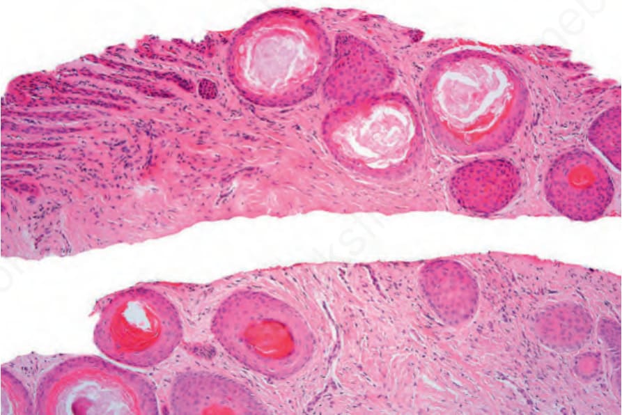
Fig. 23.47 Subungual epidermoid inclusions: numerous small inclusion cysts are present.
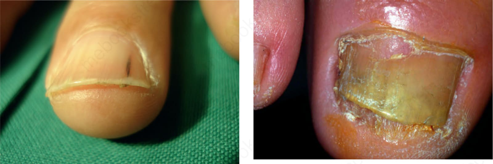
Fig. 23.50 Keratoacanthoma: the tumor is located in the proximal part of the nail apparatus. There is painful paronychia and focal nail plate destruction.
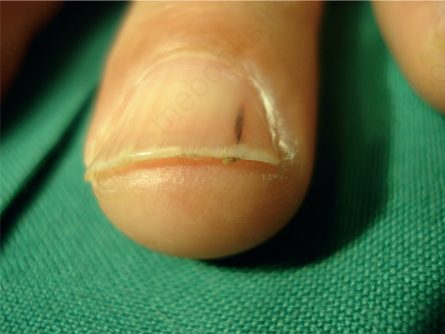
Fig. 23-48 (caption embedded in image / 圖說烘焙於圖內)