Subungual epidermoid cysts
Subungual epidermoid cysts
There are two main types of subungual epidermoid cysts1:
• Epidermoid implantation cyst is usually secondary to heavy or penetrating trauma with implantation of epidermis into the subcutaneous tissue, or even into the bone with associated osteolysis. It may be observed after inadequate wedge excision for ingrowing nails and implantation of matrix epithelium. Histopathology shows a simple epidermoid and/or ‘onycholemmal’ (matrix) cyst filled with orthokeratin and lined by a thin epithelium.2
• Subungual epidermoid inclusion cysts are mainly observed in the thumb or great toenail. Histologically, they are similar to implantation epidermoid cyst but are microscopic in size and usually an incidental histologic finding in a nail presenting with subungual hyperkeratosis and a shortened dystrophic nail plate. They have also been observed in pincer nails, clubbed nails, and even in normal nails. Subungual epidermoid inclusions frequently occur in the nail bed or distal nail matrix and result from bulbous proliferation of the rete ridges with cyst formation (Fig. 23.47). They contain homogeneous keratin without a granular layer.3 Calcification may sometimes be observed. These cysts represent a benign, probably reactive, process.1, 4, 5
Pathological features The etiology of onychopapilloma is unknown, but possible relevant factors include local trauma and/or true benign neoplastic process.2
Pathology reveals papillomatous acanthosis arising from the distal part of the matrix and spreading to the distal part of the nail bed, longitudinal canaliform deformity of the ventral nail plate, matrix-like metaplasia of the nail bed, and distal subungual hyperkeratosis (Fig. 23.49).3
Multinucleated epithelial cells with 2 to 20 nuclei may be observed. Onychopapilloma with multinucleated cells was originally described as ‘localized multinucleate distal subungual keratosis’.4
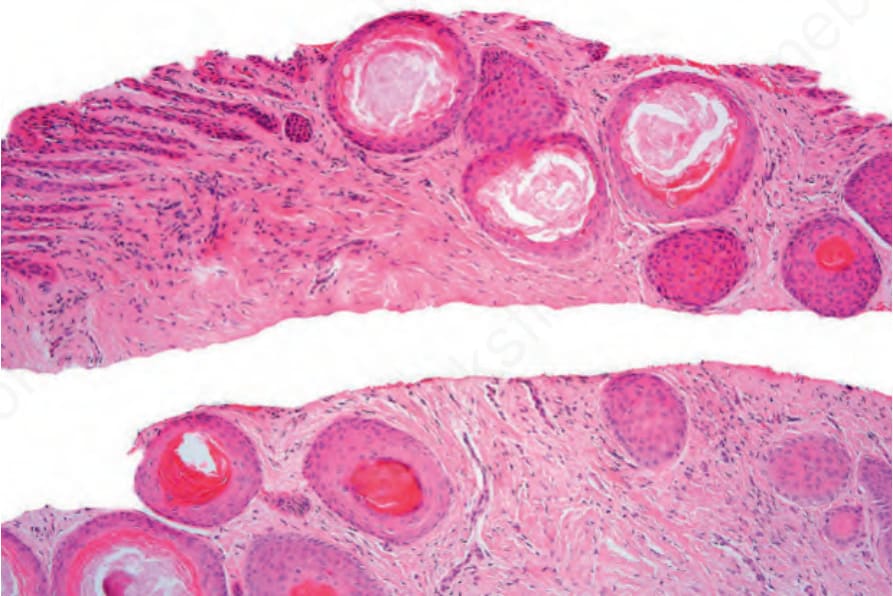
Fig. 23.47 Subungual epidermoid inclusions: numerous small inclusion cysts are present.
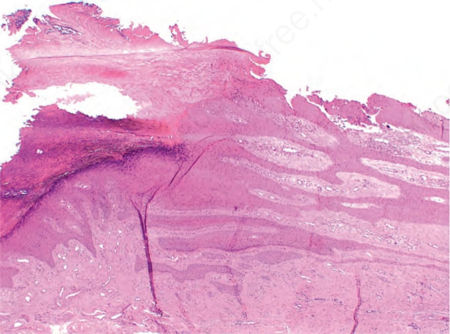
Fig. 23.49 Onychopapilloma: distal subungual hyperkeratosis and marked nail bed papillomatosis. The nail plate had been avulsed during surgery.