Longitudinal melanonychia (melanonychia striata)
Longitudinal melanonychia (melanonychia striata)
Definition Most melanocytic lesions of the nail apparatus present as a longitudinal or a total melanonychia. A longitudinal melanonychia is a longitudinal pigmented band extending from the matrix up to the distal part of the nail plate, caused by the presence of melanin in the nail plate (Fig. 23.33). In total melanonychia, the whole nail plate is pigmented. The melanosomes originate from matrix melanocytes and are transferred via their dendrites to differentiating matrix cells and will be incorporated in the nail plate. Longitudinal melanonychia may be the first sign of a nail apparatus melanoma, especially when it involves a single digit. This is why biopsies are
1140 Diseases of the nails
to perform the matrix biopsy after proximal nail avulsion and direct visualization of the pigmented area in the matrix.
Histologic examination of the nail matrix allows the identification of two broad groups of longitudinal melanonychia: melanocytic activation and melanocytic proliferation, frequently designated as melanocytic hyperplasia. Their relative incidence is completely different in adults and in children: 73% of single-digit lesions in adults are due to melanocytic activation, while 75% of single-digit lesions in children are due to benign melanocytic hyperplasia, mainly nevi.2,3
• In melanocytic activation, melanotic pigmentation of the matrix epithelium is seen, without any increase in the density of melanocytes.
• Melanocytic hyperplasia is defined as an increased number of matrix melanocytes. Benign melanocytic hyperplasia can be subdivided into lentigo when benign melanocytes remain arranged in individual units or nevus when at least one nest is present. The proliferation of melanocytes may also be malignant, corresponding to in situ or invasive melanoma. Although a number of clinical and dermatoscopic features can help in the distinction between benign melanonychia and melanoma, accurate diagnosis requires histologic examination.
Differential diagnosis A longitudinal pigmented band extending from the matrix but stopping at the distal part of the nail bed, and leaving the free edge of the nail plate unpigmented, has been described. It is due to a pigmented cornified acanthoma of the nail bed. This can be considered as the equivalent of a pigmented seborrheic keratosis.4 Melanin should not be confused with other chromogens that are not stained with the Fontana-Masson reaction. In the absence of melanin, subungual hematoma is the main differential diagnosis. It is characterized by degrading red blood cells. Perls reaction is always negative in the nail plate, as iron requires macrophage processing to be transformed into hemosiderin.
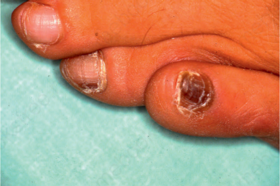
Fig. 23.33 Longitudinal melanonychia: there is involvement of the fourth toenail and complete melanonychia of the fifth toenail. This has resulted from melanocytic activation due to repeated friction from ill-fitting shoes.

Fig. 23.34 Melanocytic activation: scanning view of nail matrix.