Darier-white disease
Darier-white disease
observed with more specific changes in many nail disorders such as psoriasis and lichen planus. Typical spongiotic changes may also be associated with contact dermatitis, atopic dermatitis, and dyshidrosis. In all of these, the spongiosis usually extends to involve the periungual tissues. This is not seen in alopecia areata.
Clinical features Nails are affected in 92% of patients with Darier-White disease,1 and involvement may also be seen in the absence of any other evidence of the disease.2 The number of abnormal nails ranges from two or three, to all nails in a minority of patients.1 The fingernails are more severely affected. Characteristic features are longitudinal red and white streaks associated with distal wedge-shaped subungual keratosis.2
Histologic features Each component of the nail apparatus may be affected, but the most dramatic changes are seen in the nail bed.2,3 White longitudinal streaks and subungual keratosis are characterized by epithelial hyperplasia with marked parakeratosis and numerous multinucleated (from 2 to more than 20 nuclei) epithelial cells (Fig. 23.31). The longitudinal red streaks are due to mild epithelial hyperplasia and vasodilatation. The histologic findings in the nail bed of Darier disease differ from those of the skin: presence of multinucleate epithelial giant cells and near-absence of inflammatory infiltrate. The matrix may be completely spared. However, if the distal nail matrix is affected, typical changes of Darier disease may be observed. The proximal matrix is rarely affected.
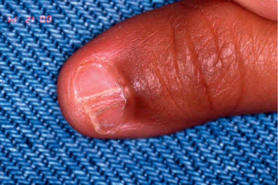
Fig. 23.30 Lichen striatus: there is a median, longitudinal, lichenoid nail involvement. Discrete flesh-colored linear papules are present on the dorsum of the finger. The patient is a 2-year-old boy.
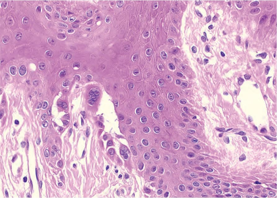
Fig. 23.31 Darier-White disease: suprabasal acantholysis with multinucleated cells.