Psoriasis
Psoriasis
Clinical features Nail involvement is frequent in psoriasis, occurring in 10% to 50% of patients.1 It is estimated that over a lifetime, between 80% and 90% of psoriatic patients will suffer nail disease.2 Fingernails are more frequently affected than toenails. Several nails are usually affected, but involvement of a single nail can sometimes be seen.
The clinical appearance depends upon the part of the nail involved. Matrix involvement results in pitting, leukonychia, nail plate thickening or thinning, onychorrhexis, and crumbling (Fig. 23.19). Nail bed involvement gives rise to the ‘oil drop’ or ‘salmon patch’ signs, splinter hemorrhages, subungual hyperkeratosis, and onycholysis. PNF involvement can mimic chronic paronychia. Pitting, onycholysis, discoloration, and subungual hyperkeratosis are the most common symptoms.1,3,4
Pustular psoriasis of the nail, also known as Hallopeau acrodermatitis continua, is mainly observed in middle-aged females and has a chronic, relapsing course. It generally affects a single digit and is usually not associated with other manifestations of pustular psoriasis. Involvement of the nail bed with pustules, scale crusts, and onycholysis is seen.5,6
Parakeratosis pustulosa typically affects a single fingernail (thumb or index) in girls younger than 7 years. It is a chronic condition not regarded as a specific disease but as a manifestation of atopic dermatitis, contact dermatitis, and psoriasis. It is characterized by erythematosquamous paronychia accompanied by intermittent vesicles and pustules, onycholysis, mild distal or lateral hyperkeratosis, and nail plate deformities.7–9 Although the disease generally resolves after a few years, some children develop psoriasis.9
In pustular psoriasis, true spongiform pustules can be observed in the nail bed. They may coalesce to form intraepidermal or subcorneal macropustules (Figs 23.21 and 23.22).5
In parakeratosis pustulosa, histology may show spongiotic or psoriasiform changes.9,17
Differential diagnosis A PAS stain should always be performed to exclude onychomycosis because the latter may clinically and histologically mimic nail psoriasis and because a mycotic superinfection or colonization is observed in 13% to 27% of cases of nail psoriasis.18,19
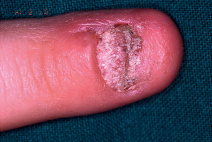
Fig. 23.19 Psoriasis: note the scar from the prior lateral-longitudinal biopsy. The nail plate is dystrophic and thickened. Courtesy of B. Richert, MD, PhD, Université Libre de Bruxelles, Belgium.
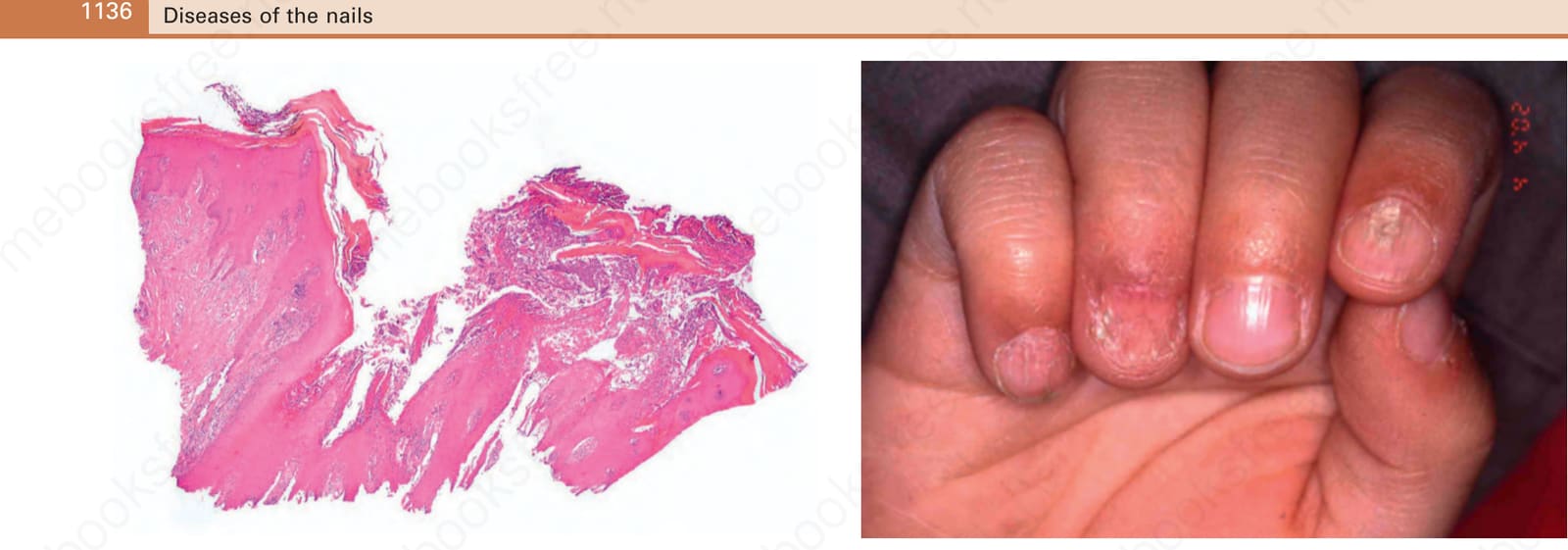
Fig. 23.21 Pustular psoriasis: there is crusting, parakeratosis, and a macropustule.