Clinical features2
Clinical features2
Periungual warts are frequently seen in children and young adults. They are mainly located around the nail, leaving the nail plate generally unaffected. They present as firm, keratotic papules with sizes ranging from a few millimeters to several centimeters in diameter. Subungual warts generally involve the hyponychium, located in the distal nail bed, and cause subungual hyperkeratosis, or onycholysis. Presentation under the PNF is rare, although the clinical appearance is typical, with a paronychia-like inflammatory reaction and distal wart papillae emerging from under the cuticle (Fig. 23.18).3
The diagnosis of a periungual wart is generally made from the clinical appearances. However, resistant periungual warts in adults require a biopsy, particularly when only one finger is involved, in order to exclude Bowen disease or an amelanotic melanoma.
Pathogenesis and histologic features Periungual warts are benign epithelial tumors caused by the human papillomavirus (HPV), usually HPV 2 and 4. The histologic appearances include acanthosis, papillomatosis, parakeratosis, and koilocytes in the most superficial layers. In cases of HPV 1-induced infection (myrmecia), the keratinocytes contain numerous inclusions resembling eosinophilic coarse-grained keratohyaline.
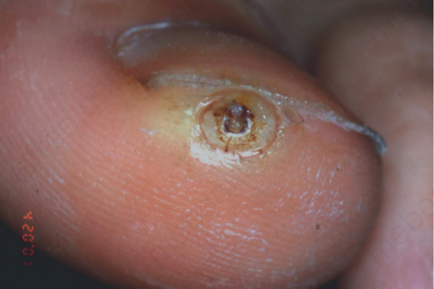
Fig. 23.16 Tungiasis: a typical site. The flea is partially visible.
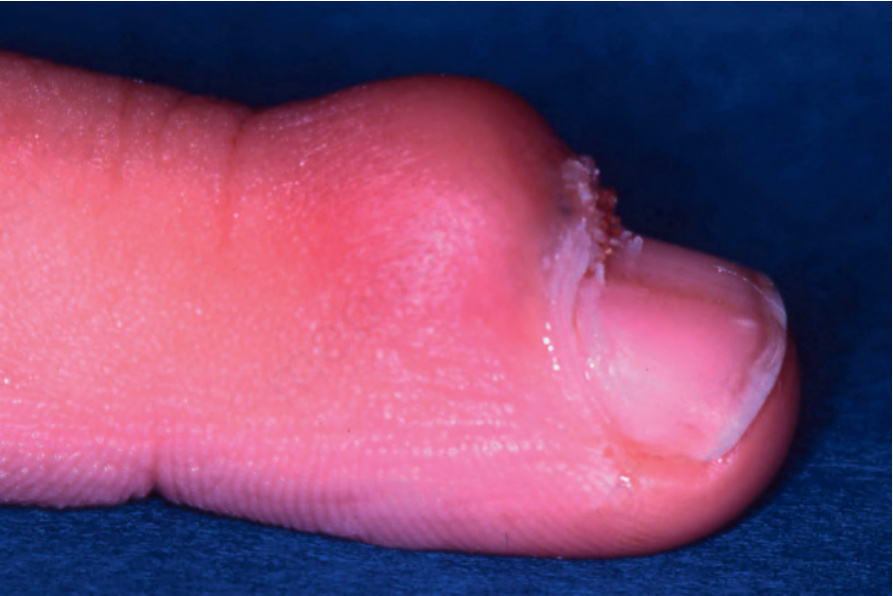
Fig. 23.18 Viral wart: note the white papillary processes just protruding at the proximal nail fold.