Trichostasis spinulosa
Trichostasis spinulosa
Clinical features Trichostasis spinulosa is a very frequent alteration of the hair follicle characterized by dilated vellus hair follicles with retention of successive telogen hairs that protrude from a single dilated ostium. The disorder was first recognized by Felix Franke in 1901, who named it ‘Das Pinselhaar Thysonatrix’ (paintbrush hair). The term trichostasis spinulosa was coined by Noble in 1913.1
Lesions present as multiple hyperkeratotic follicular papules that usually resemble comedones, but close examination may reveal a tuft of hairs. It has predilection for areas with abundant pilosebaceous units, including the face, neck, chest, upper arms, and interscapular area (Fig. 22.184). The defect is seen after adolescence in males and females, but it is more commonly seen in elderly patients.
There are two variants: a classic nonpruritic form that presents on the face (particularly on the nose of middle-aged to elderly individuals as a solitary comedo-like lesion) and a pruritic variant that especially involves the limbs and trunk of young adults.2–4 It has also been classified as primary trichostasis spinulosa when it is seen as an isolated finding or as secondary trichostasis spinulosa when it is described within pre-existing skin lesions.
Differential diagnosis Conditions presenting as keratotic follicular papules may be considered in the differential diagnosis. These include keratosis pilaris, ichthyosis follicularis, hypovitaminosis A and C, eruptive vellus hair cysts, and Darier disease. It also has to be differentiated from entities such as comedonal acne and Favre-Racouchot syndrome.1
Pathogenesis and histologic features The pathogenesis is unknown although some studies have suggested that Pityrosporum and/or P. acnes may be responsible for induction of follicular hyperkeratosis with vellus hair retention.5 Follicles may also contain Malassezia furfur yeasts. Difusse trichostasis spinulosa has been reported in prolonged use of topical corticosteroids and chronic renal failure.1,6
Histology reveals a dilated hair follicle in which a keratin plug surrounds multiple hair shafts derived from a single matrix and papilla (Fig. 22.185). Dermoscopy may help identify the characteristic hair tuft.7
Trichostasis spinulosa has been described as an associated finding in intradermal melanocytic nevi, seborrheic keratoses, syringomas, and nodular basal cell carcinomas.8–11
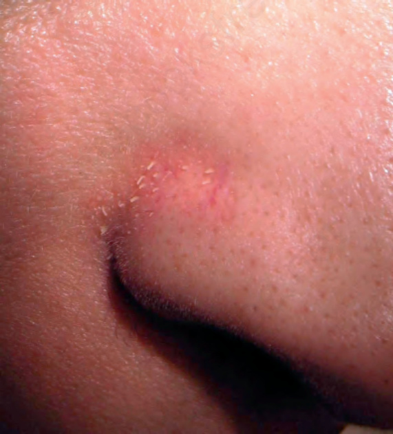
Fig. 22.184 Trichostasis spinulosa: note the follicular prominence on the nasal fold. Courtesy of L.M. Gómez, MD, UPB, Medellín, Colombia.
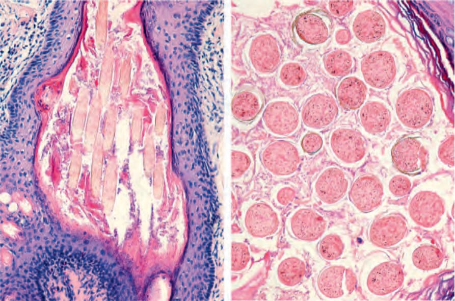
Fig. 22.185 Trichostasis spinulosa: vertical and horizontal sections. Numerous nonpigmented hair shafts in a single hair follicle.