Pili multigemini
Pili multigemini
Clinical features Pili multigemini (multi hair) was first described by Flemming in 1883 and Giovanni in 1892. It is a hair shaft developmental abnormality, which occurs mostly on the face, particularly the beard area and the scalp in children, but has also been described with involvement of the entire body. Several hair shafts emerge from a single follicular ostium.1
Under light microscopy, the hair shafts may initially seem normal but when horizontal sections are examined the hair follicles and shafts are triangular, kidney- or heart-shaped. The hair shafts may also have a longitudinal canalicular dent or a prominent longitudinal groove along the long axis of the hair (Fig. 22.181). In some cases, the internal root sheath forms an angle which results in hair follicle deformity. The best method to observe the triangular hair configuration and canalicular dent is with scanning electron microscopy. More than 50% of the hairs from an individual with this syndrome will have this characteristic appearance, compared to less than 5% in the general population.4,16–18
Differential diagnosis The differential diagnosis includes pili torti, monilethrix, woolly hair, and acquired progressive kinking of the hair. Overall, the clinical features are typical and the differential diagnosis is usually straightforward.19
The condition is usually asymptomatic, but it may be associated with erythema and recurrent follicular inflammation leading to scarring. In some cases, the abnormality follows Blaschko lines. An association with cleidocranial dysostosis has been documented.2,3
Most cases of pili multigemini have been found incidentally. A recent study showed that all the Europeans randomly chosen had the condition. Because of this finding, it has been concluded that, rather than a rare development defect of hair, it should be considered as a varying vestige of evolution.4
Histologic features Pili multigemini is characterized by several hair shafts within a single follicular canal (from two to eight), each of which originates from an individual matrix and papilla (Fig. 22.183). The hair may have a flattened or
1116 Diseases of the hair
triangular shape and has all the components of a normal follicle except that there is a common external root sheath.2
Differential diagnosis The condition must be distinguished from tufted folliculitis. In the latter condition, fusion of hair follicles occurs in the upper segment of the follicle as a result of inflammation, and each hair is surrounded by its own external root sheath.
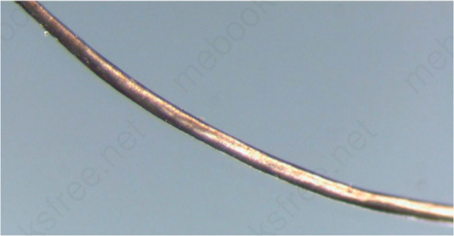
Fig. 22.181 Uncombable hair syndrome (pili canaliculi): there is a longitudinal grooving within the hair (pili canaliculi) which contributes to the structural rigidity and clinical appearance. Courtesy of A.M. Aristizábal, MD, CES, Medellín, Colombia.
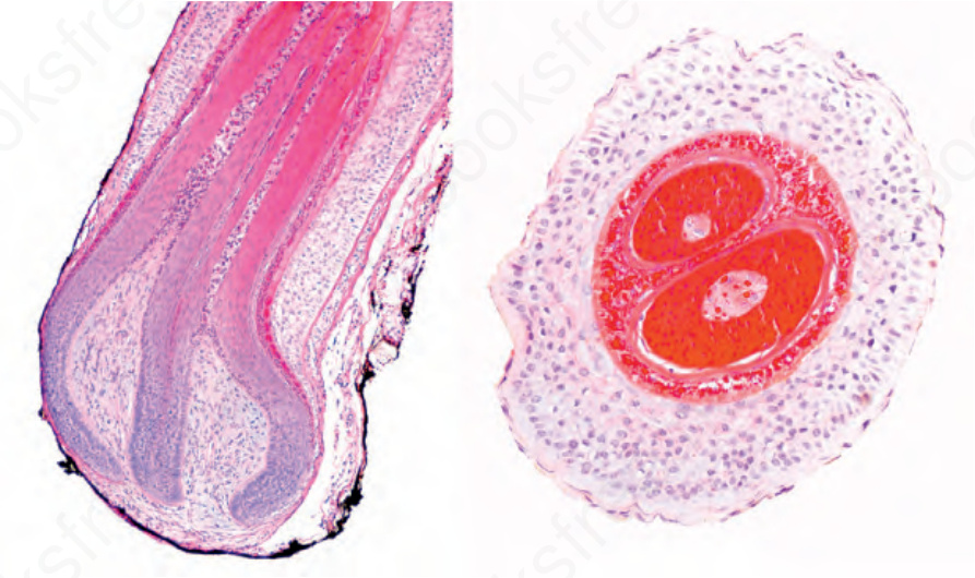
Fig. 22.183 Pili multigemini: in these vertical and horizontal sections, note the two hair shafts within a single follicular canal. The hairs have all the components of a normal follicle and a common external root sheath.
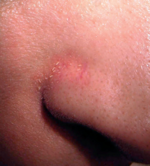
Fig. 22.184 Trichostasis spinulosa: note the follicular prominence on the nasal fold. Courtesy of L.M. Gómez, MD, UPB, Medellín, Colombia.