Riehl melanosis
Riehl melanosis
Clinical features Riehl melanosis, first described in 1917, consists of prominent hyperpigmentation of the face with involvement of the forehead and the zygomatic and/or temporal regions (Fig. 20.49).1 The condition has been reported in both Caucasians and patients with darker skin. Women are mainly affected.
Pathogenesis and histologic features Riehl proposed that the changes were secondary to some food items used during the First World War. However, more recently, the condition has been regarded as a variant of pigment contact dermatitis. The most common allergens involved are cosmetics, including fragrances.2 Riehl melanosis has rarely been reported in association with Sjögren syndrome and lichen planus.3,4
Histologically, two histopathologic patterns have been described: epidermal and dermal. In the former, there is increase in pigmentation of basal cells within the epidermis. In the latter, there is pigmentation in the upper dermis with melanophages and decreased pigmentation in basal cells.10 Interestingly, a study on exogenous hyperpigmentation has found an increase in the number of basal melanocytes in affected areas as compared to normal skin.11 In some cases, colloid bodies are seen at the dermal–epidermal junction suggesting a pre-existing interphase tissue reaction.
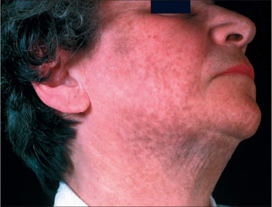
Fig. 20.49 Riehl melanosis: prominent patchy hyperpigmentation of the face. By courtesy of the Institute of Dermatology, London, UK.