Carney complex
Carney complex
Clinical features This syndrome was reported at the beginning of the 1980s under the acronyms NAME (nevi, atrial myxomas, myxoid neurofibromas and ephelides) and LAMB (lentigines, atrial myxoma, mucocutaneous myxomas and blue nevi) and was delineated in 1985 by Carney in a large series of patients.1–3
1010 Disorders of pigmentation
Skin involvement was the predominant finding in four generations of a reported kindred and it has been suggested that, in these cases, the presence of a pilonidal sinus is an associated feature of the disease.35
Parenchymal breast lesions include myxoid fibroadenoma and (rarely) ductal adenoma.36,37
Pathogenesis and histologic features Carney complex is a genetically heterogeneous disease. So far, the genes responsible for the syndrome have been mapped to chromosomes 17q22-q24 and 2p16.38–41 In up to 65% of the affected patients investigated there were heterozygous inactivating mutations in the tumor suppressor gene PRKAR1A that maps to 17q and codes for the type Iα regulatory subunit of protein kinase A. This protein is an essential component of many cellular signaling systems and it appears to be important in cardiac function and myxogenesis.42 A pituitary-specific knockout of the PRKAR1A model in mice induced tumors in the gland.43 Tumors from affected individuals have shown decreased basal activity of protein kinase A but an increase in the cAMP-stimulated activity, which may lead to tumorigenesis.44–46 Missense mutations in PRKAR1A are very unusual.47 Patients with no PRKAR1A gene defects identified by sequencing may have large PRKAR1A deletions in the germline.48
The cardiac myxomas have features that are indistinguishable from other cardiac myxomas and present as sessile or polypoid mobile masses. Histologically, there is an abundant myxoid matrix, variable vascularity, and scattered tumor cells. These cells are bland and may be elongated or stellate. Exceptionally, ossification and extramedullary hematopoiesis have been reported.49
Endocrine and testicular tumors are identical to their counterparts occurring in a nonfamilial setting and will not be described here.
Cutaneous myxomas are identical to sporadic cutaneous angiomyxomas except for the fact that the latter tend to be more ill defined. The tumor is well circumscribed and hypocellular, with prominent myxoid change and abundant small thin-walled vascular channels (Fig. 20.41).7 The cells within the lesion are bland, short, spindle-shaped or stellate, with vesicular nuclei (Fig. 20.42). Mitotic figures are rare. An epithelial component – probably
1011 LEOPARD syndrome
Lesions previously described as psammomatous melanotic schwannoma are now classified along with the identical sporadic counterpart as malignant melanotic schwannian tumors as they all have potential for malignant behavior regardless of the histologic features.51 These tumors are very rare in the skin and tend to be fairly circumscribed but not encapsulated containing a mixture of spindled and epithelioid melanocytes with variable pigmentation and often, but not always, psammoma bodies (Fig. 20.44). The bundles of melanocytes may show whorling and there is focal nuclear palisading. Cytological atypia may be mild and prediction of behavior cannot be made based on histologic features.16,17,52
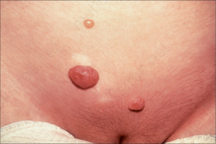
Fig. 20.37 Carney complex: multiple soft tumor nodules are evident on the lower trunk. By courtesy of M. Walsh, MD, Royal Victoria Hospital, Belfast, UK.
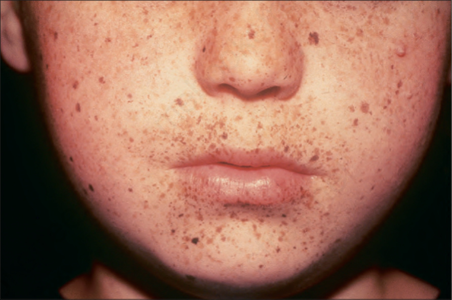
Fig. 20.38 Carney complex: numerous lentigines on the central face. By courtesy of M. Walsh, MD, Royal Victoria Hospital, Belfast, UK.
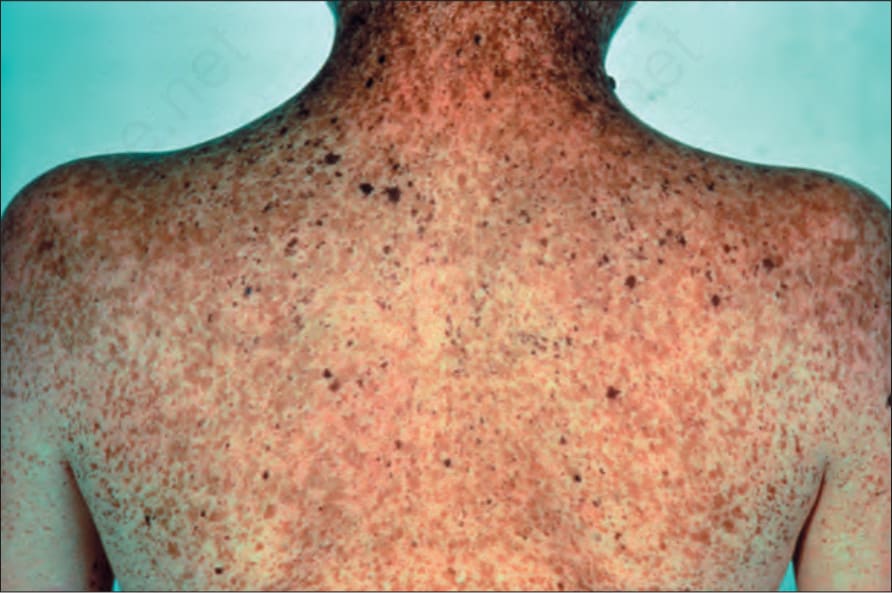
Fig. 20.39 Carney complex: multiple lentigines and scattered banal nevi and blue nevi on the trunk. By courtesy of D. Atherton, MD, St John’s Institute of Dermatology, London, UK.
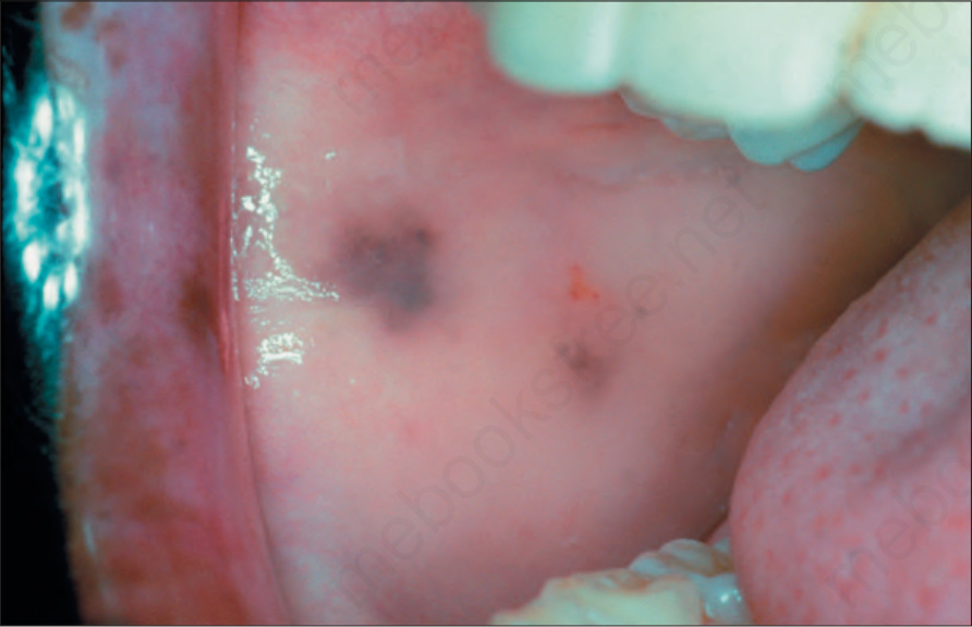
Fig. 20.40 Carney complex: pigmented macules in the oral cavity are rare. By courtesy of the Institute of Dermatology, London, UK.
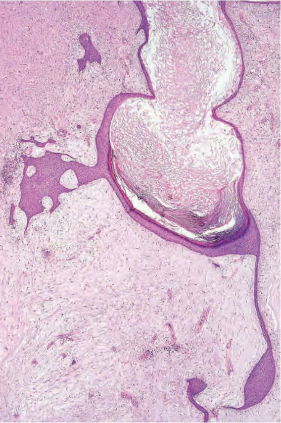
Fig. 20.41 Cutaneous angiomyxoma: dermal poorly cellular angiomyxoma with cystically dilated follicular structure.
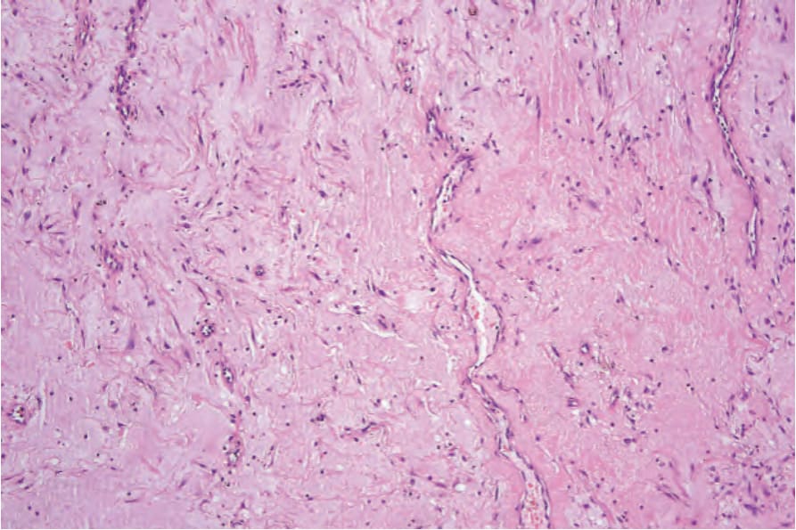
Fig. 20.42 Cutaneous angiomyxoma: bland stellate and short spindle-shaped cells in a myxoid stroma with prominent small blood vessels.
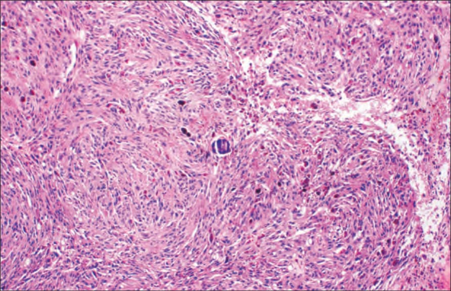
Fig. 20.44 Psammomatous melanotic schwannoma: pigmented spindle-shaped cells. There is a psammoma body in the center.