Peutz-Jeghers syndrome
Peutz-Jeghers syndrome
Clinical features Peutz-Jeghers syndrome is an autosomal dominant disease characterized by gastrointestinal polyposis and pigmented macules with involvement of perioral skin, lips, buccal mucosa, and hands and feet – particularly palms, soles, fingers, and toes (Figs 20.35 and 20.36).1–3 They may rarely occur on the nose and cheeks. Pigmentation of the umbilicus has been reported in one case.4 Lesions are brown or black and usually measure less than 5 mm in diameter, although acral lesions may be larger. Cutaneous macules can be congenital or appear very early in life, usually before the age of 2 years. Skin lesions may fade at puberty but mucosal lesions tend to persist. The clinical appearance is almost identical to that of lentigines. Interestingly, pigmented macules have been reported in psoriatic plaques.5,6 Males and females are equally affected. Patients presenting without a family history are likely to have developed the disease as a result of a spontaneous mutation.
Pathogenesis and histologic features The disease has been cloned to chromosome 19p13.3. It is caused by germline mutations in LKB1 (STK11) responsible for encoding a serine/threonine kinase that acts as a tumor suppressor gene.10–12 Mutations in LKB1 are found in up to 80% of patients with the disease.13 Rare cases have been cloned to chromosome 19p13.4.14 The LKB1 protein is present in both the nuclei and the cytoplasm and translocates to the mitochondria during apoptosis.15 It associates with p53 and regulates specific p53-dependent apoptosis.15 The gastrointestinal polyps in Peutz-Jeghers syndrome do not stain for LKB1 whereas this protein is highly expressed in pyknotic nuclei of neighboring intestinal cells. This suggests that the polyps are formed as a result of a deficiency in apoptosis and may also explain the development of cancer in this syndrome.15 It has been suggested that the LKB1 mutations lead to the activation of the Wnt/beta-catenin pathway and this is a contributor to the neoplastic predisposition in the syndrome.16,17 Interestingly, in some families with Peutz-Jeghers syndrome germline mutations in LKB1 are not found, suggesting that mutations in an unidentified gene may cause the disease in this group of patients.18,19
Identification of cutaneous lesions is important because they usually precede the clinical manifestations of the gastrointestinal polyps. The polyps seem to have low potential for malignant transformation. They are
Histologically, the skin lesions show increased pigmentation of basal cells but there appears to be no increase in the number of basal melanocytes. The latter allows distinction from a lentigo. Histologic distinction from a freckle is not possible.
The intestinal polyps do not have any specific features that allow distinction from other polyps. In some cases, misplacement of the epithelium in the polyps into the submucosa, muscularis propria, or subserosa has been documented and this may be confused with malignancy.20
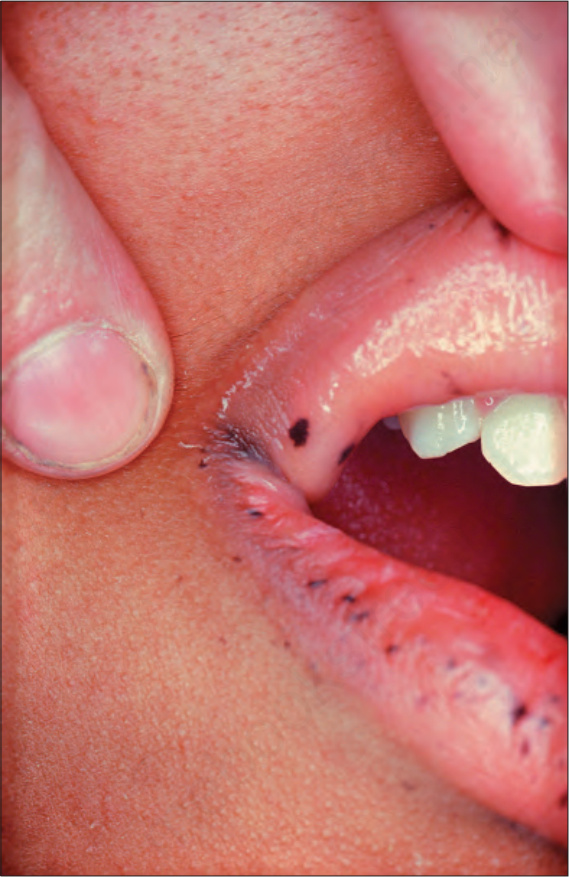
Fig. 20.35 Peutz-Jeghers syndrome: darkly pigmented macular lesion on the lips. By courtesy of the Institute of Dermatology, London, UK.
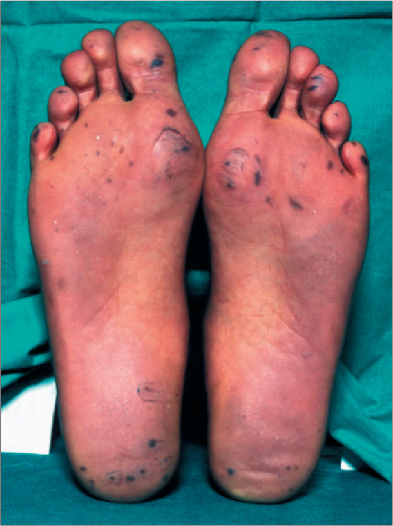
Fig. 20.36 Peutz-Jeghers syndrome: multiple darkly pigmented larger macular lesions on the soles. By courtesy of the Institute of Dermatology, London, UK.