Laugier-Hunziker syndrome
Laugier-Hunziker syndrome
Clinical features Laugier-Hunziker syndrome (idiopathic lenticular mucocutaneous pigmentation) is an acquired disorder characterized by macular pigmentation of the lips, oral cavity (mainly hard palate and less commonly buccal mucosa, soft palate, and gums), and, less frequently, fingers and palms (Fig. 20.24).1–6 Longitudinal melanonychia is seen in up to 50% of patients.1,7 Pigmentation of the proximal nail fold resulting in a pseudo-Hutchinson sign may be seen. Genital and conjunctival pigmentation is very rare (Fig. 20.25).4,8 The sex incidence is similar and most patients are Caucasian and middle aged. The pigmented macules are not associated with any systemic disease. Familial occurrence is exceptional.9 A case with esophageal melanocytosis has been documented.10 A patient with coexistent actinic lichen planus has also been reported.11 In a further patient, macular pigmentation developed at the sites of irritant contact dermatitis and viral warts.12 Hyperpigmentation identical to that seen in Laugier-Hunziker syndrome may occur in association with chemotherapy for cancer and, in one case, after levodopa therapy.13,14 A melanoma has been reported in a mucosal lesion.15
Histologically, two main patterns have been described: epidermal and mixed. In the former, there is an increase in the content of melanin in keratinocytes in all levels of the epidermis.20–24 In the latter, there is pigmentation
The dermoscopic appearance of the pigmented lesions has been described.16–18 Macular pigmented lesions on oral mucosa and genital skin show a parallel pattern; lesions of melanonychia show longitudinal brown and gray lines and bands with ill-defined margins in the lesions of melanonychia. Acral macular lesions show a parallel furrow pattern.
Pathogenesis and histologic features The pathogenesis of the pigmentation is not yet known.
Histologically, there is hyperpigmentation of basal keratinocytes and pigment incontinence with melanophages in the papillary dermis. Recently, it has been suggested that there is an increase in the number of basal dendritic melanocytes and the term ‘mucocutaneous lentiginosis’ of Laugier and Hunziker has been proposed.19
Ultrastructural studies show the presence of increased mature melanosomes in the cytoplasm of keratinocytes.2
Differential diagnosis Distinction from Peutz-Jeghers syndrome cannot be made based on histologic examination alone, and close clinicopathological correlation is therefore required.20 Primary adrenocortical insufficiency may clinically simulate Laugier-Hunziker syndrome.21
1003 McCune-Albright syndrome
A
B
C
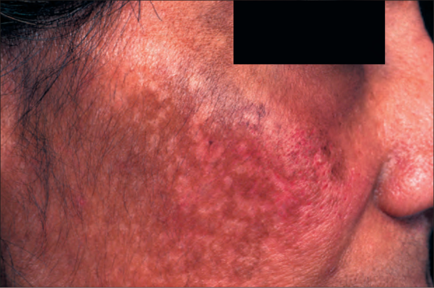
Fig. 20.23 Melasma: dark-brown macular pigmentation. By courtesy of the Institute of Dermatology, London, UK.
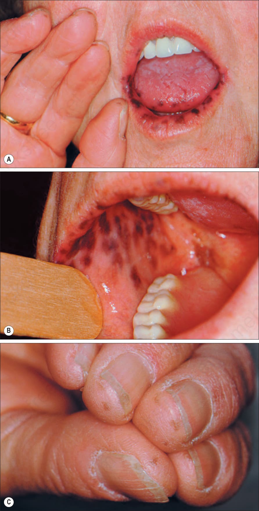
Fig. 20.24 Laugier-Hunziker syndrome: pigmented macules on (A) the lips, (B) the buccal mucosa, and (C) the fingers. By courtesy of I. Viana, MD, Lisbon, Portugal.
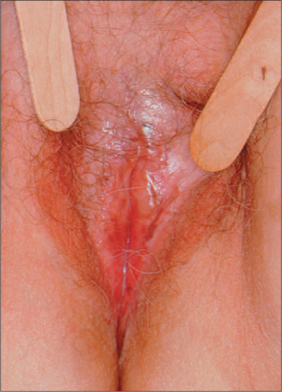
Fig. 20.25 Laugier-Hunziker syndrome: pigmented macules on the vulva. By courtesy of I. Viana, MD, Lisbon, Portugal.