Nevus depigmentosus
Nevus depigmentosus
Clinical features Nevus depigmentosus (also known as nevus achromicus) is a common nevoid abnormality present in 0.5% to 1.25% of neonates or develops in the first few years of life.1–3 A single, well-defined, macular, rounded hypopigmented patch, ranging in size from less than 1 cm to several centimeters, is characteristically seen.4,5 In rare cases the distribution may be segmental or systematized along Blaschko lines (Fig. 20.19).6–8 In the latter setting the lesion resembles hypomelanosis of Ito. There is predilection for the trunk and proximal extremities. In one case, involvement of the iris was seen.9 Rare associations include pes cavus, mental retardation, hemihypertrophy, inflammatory linear verrucous epidermal nevus, lentiginosis (unilateral or segmental), congenital linear punctate keratoderma, Becker nevus, nevus flammeus, nevus spilus, and eccrine angiomatous nevus.6,10–17 In rare cases, melanocytic nevi or lentigines may develop within lesions of nevus depigmentosus, in one case lentigines developed after UVB therapy, and in a further case a nevus depigmentosus with lentigines was associated with underlying breast hypoplasia.18–23
Pathogenesis and histologic features Mutations in the PAX-3 gene located on chromosome 2q37 have been reported in Waardenburg syndrome types I and III.9–11 Mutations in the MITF gene located on chromosome 3p14.1-p12.3 and SOX10 are responsible for about 30% of cases of the cases of Waardenburg syndrome type II.12,13 EDNRB (endothelin receptor type B) mutations are found in the heterozygous state in between 5% to 6% of cases. In the rest, a specific mutation has not been identified.13,14 Type IV syndrome is associated with mutations in the endothelin 3 gene and in the endothelin receptor type 3.15 Cases with neurological disease also show mutations in SOX10 and the latter has been associated with type II syndrome with or without neurological symptoms.6,16
Histologically, the patches of hypopigmentation in all variants of the disease show absence or reduction in the number of epidermal melanocytes.
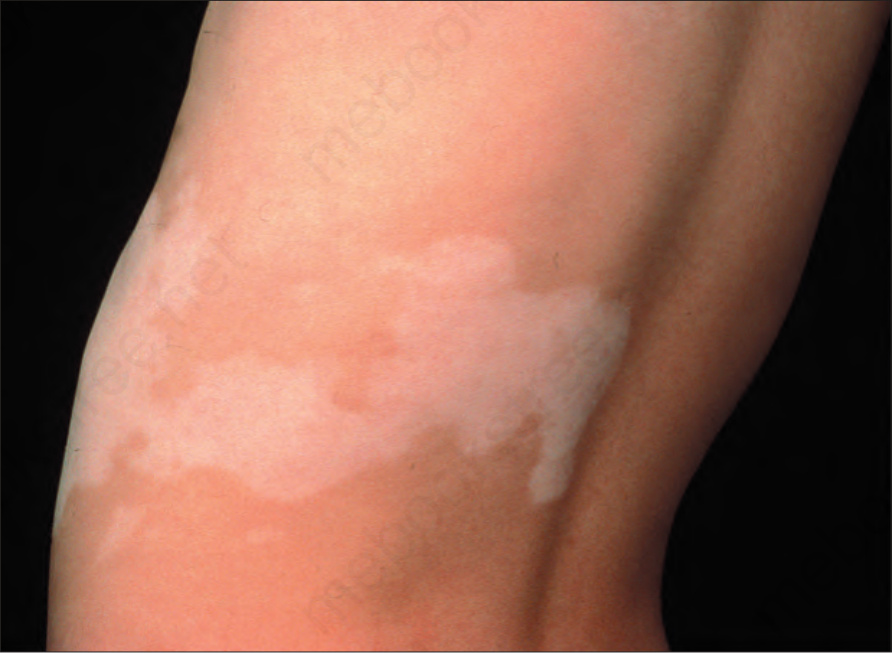
Fig. 20.19 Nevus depigmentosus: segmental distribution. By courtesy of the Institute of Dermatology, London, UK.