Piebaldism
Piebaldism
Clinical features Piebaldism is a rare congenital disorder inherited in an autosomal dominant manner. In the past, the name ‘partial albinism’ was used as a synonym but this term is no longer accepted, as piebaldism is not considered to be a variant of albinism.1 It is characterized by localized macular areas of hypopigmentation and affects all races, with the same sex incidence.2,3 The central area of the forehead, the anterior trunk, and the mid areas of the extremities are characteristically affected (Figs 20.16 and 20.17). There may be involvement of the eyelashes and of the medial aspect of the eyebrows but the hands, feet, and back are usually spared. In 90% of cases there is a white forelock in the frontal mid scalp (Fig. 20.18); this may be the only manifestation of the disease. Poliosis circumscripta refers the absence of pigment in a circumscribed hairy area and, although more frequently seen on the scalp, it can affect many areas of the body, including the eyebrows and eyelashes. Poliosis is not specific of piebaldism and can be associated with other genetic conditions, including Waardenburg syndrome, neoplastic conditions, and even medications. The macular areas of hypopigmentation vary in size from 1 cm to several centimeters. They are usually stable and show no progression, but rarely contraction, expansion of lesions, or exceptionally complete regression has been documented.4,5 Areas of hyperpigmentation may be seen either within the patches of hypopigmentation or in normally pigmented skin. Café-au-lait spots are a frequent finding and intertriginous freckling may also be seen. An association with neurofibromatosis
associated with truncated mutations in the tyrosine kinase domain resulting in haploinsufficiency or a dominant-negative effect.24
In some patients in whom KIT mutations are not demonstrated, deletion of the SLUG gene, mapped to chromosome 8, has been documented.25 SLUG is a zinc-finger neural crest transcription factor, which is critical for the development of hematopoietic stem cells, melanoblasts, and germ cells in the mouse.
Histologic examination of affected skin usually shows complete absence of melanocytes, and this is confirmed by ultrastructural studies.1,26 Abnormal melanocytes may be seen in areas of transition between involved and normal skin.
999 Nevus depigmentosus
trunk.1–3 Some cases involve the face or extremities. The area of hypopigmented skin usually measures several centimeters and may contain small patches of normal skin. A case of generalized nevus anemicus has been documented.4 Nevus anemicus is often associated with port-wine stain, nevus spilus, lymphedema, phakomatosis pigmentovascularis, tuberous sclerosis, and capillary malformation-arteriovenous malformation syndrome.5–10 It has also been recently reported as a newly noted manifestation of neurofibromatosis type 1.11,12 Lesions are more frequently noted in younger patients.13 Nevus oligemicus has been described as a variant of nevus anemicus.14,15
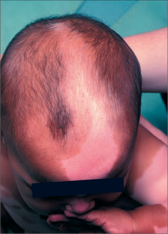
Fig. 20.16 Piebaldism: patchy hypopigmentation of the scalp. By courtesy of the Institute of Dermatology, London, UK.
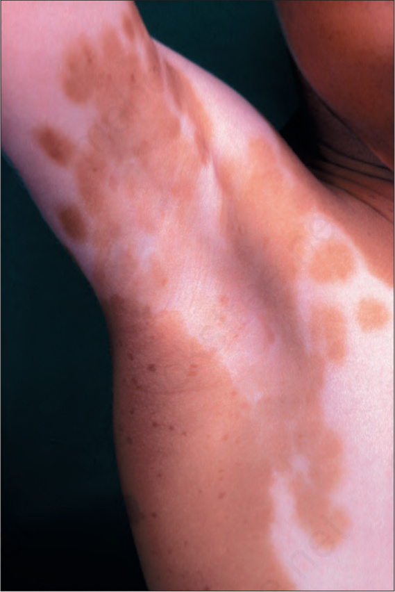
Fig. 20.17 Piebaldism: hypopigmentation of the trunk and proximal limbs. By courtesy of the Institute of Dermatology, London, UK.
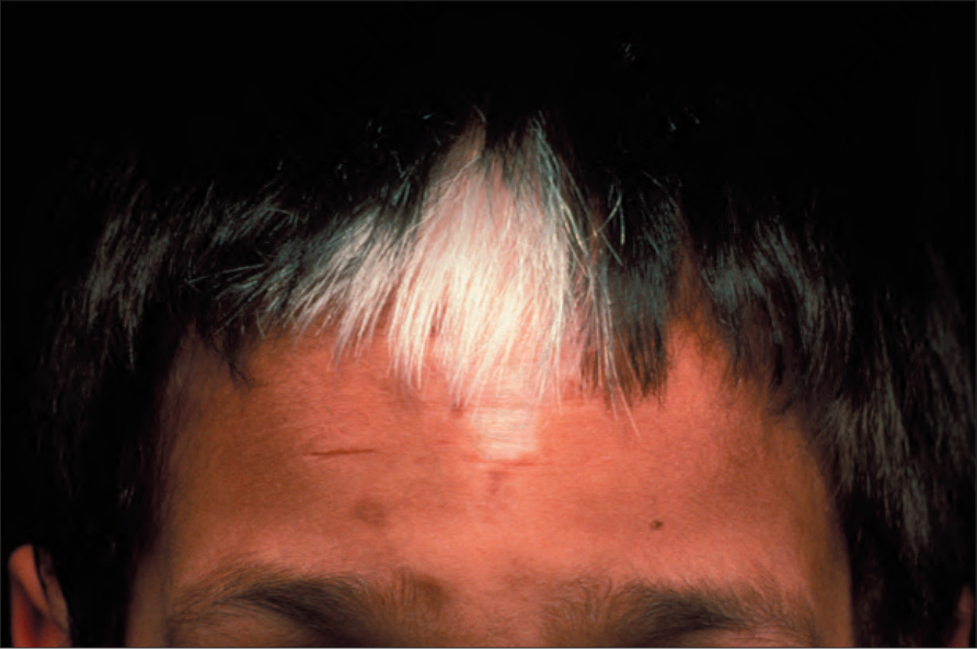
Fig. 20.18 Piebaldism: typical white forelock. By courtesy of the Institute of Dermatology, London, UK.