Alternariosis
Alternariosis
Clinical features Alternaria is a pigmented fungus within the heterogenous phaeohyphomycete group.1,2 It is associated with inoculation during minor trauma and is often found in immunocompromised hosts, especially iatrogenically immunosuppressed organ transplant recipients.3,4 Other reported clinical settings include underlying hematological malignancy, systemic corticosteroid therapy, Cushing disease, and AIDS.5 Immunocompetent individuals are affected rarely.5,6
pigmented septate hyphae measuring 5–7 µm in diameter, accompanied by open, rounded bodies 3–10 µm in diameter (range 5–20 µm) (Fig. 18.358).5 They may be seen free or in histiocytes or giant cells. There is a dermal inflammatory infiltrate usually consisting of a mixed neutrophil and granulomatous reaction. A variable number of microabscesses may be present. Although epidermal changes are often not marked, pseudoepitheliomatous hyperplasia is encountered in some cases.
Patients present with ulcers, erythematous macules and papules, pustules, and nodules on exposed skin surfaces, sometimes crusted or verrucous (Fig. 18.357).4,7 Large ulcerated plaques with pustules may also occur.8,9 Lesions may be found on the dorsa of the hands, the fingers, elbows, knees, face, and dorsa of the feet.10 A sporotrichoid distribution of skin lesions has been reported.11 The lesions occasionally regress spontaneously; otherwise, they respond well to antimycotic drugs and leave only slight scars.
Pathogenesis and histologic features Alternaria spp. are found widely in soil and plants and are most often inoculated into skin with wood splinters. In the tissues, the fungus is seen as
Differential diagnosis Alternariosis may be readily confused with blastomycosis, especially when pigmented hyphal forms are absent from the tissue sections. Culture of the organism and/or PCR studies, however, should facilitate a more definitive diagnosis.5
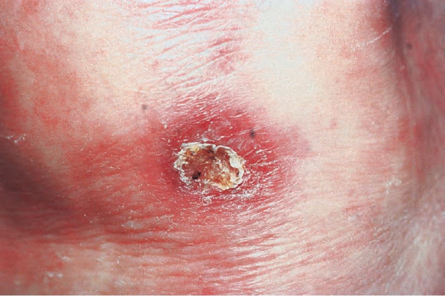
Fig. 18.357 Cutaneous alternariosis: crusted ulcer at the base of the thumb. By courtesy of S.W. Lanigan, MD, Bridgend General Hospital, Bridgend, UK.
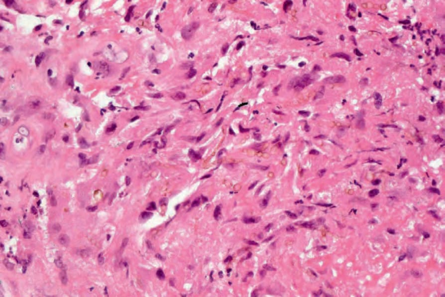
Fig. 18.358 Cutaneous alternariosis: note the yeast forms in this hematoxylin and eosin stained section.