Endothrix infections
Endothrix infections
Children are more commonly affected. Kerion is seen as an area of inflamed alopecia in which the broken-off hairs are loose in their follicles and are associated with suppuration (Figs 18.264 and 18.265). This may be severe enough to discharge via sinuses, with the formation of fibrinopurulent crusts around adjacent hairs. Secondary bacterial infection (e.g., with S. aureus) may play a part.3 Fluorescence is not a feature. The condition may be confused clinically with folliculitis decalvans or dissecting cellulitis of the scalp.3,9,10 Involvement of the beard area (tinea barbae, tinea sycosis), which occurs most often in farm workers, invariably affects adult males.11,12 The lesions appear as erythematous areas of pustular folliculitis in the beard area and may present as a kerion (Fig. 18.266).
Clinical features These infections, most often with Trichophyton tonsurans and T. violaceum, usually cause patchy alopecia with little inflammation. T. tonsurans is now the most common cause of scalp ringworm in the United States.1–4 This organism was found to be responsible for around 22% of cases of tinea capitis in a study from Nigeria.5 T. violaceum is the most common cause of tinea capitis in South Africa.6 The hair break is at the ostium of the follicle so that broken hairs are seen as dots rather than stumps. The intervening skin usually shows only slight scaling, but occasionally pustules and kerion can develop.7 Drainage lymphadenopathy may be evident. The hairs do not fluoresce with a Wood lamp.
Histologic features The hyphae of T. tonsurans and T. violaceum extend within the hair shaft and produce spores. The epidermis is patchily parakeratotic, and there are
927 Tinea capitis
A
B
remnants of infected hair shafts in the dilated keratin-plugged ostia. Perifollicular inflammation is variable in intensity, but often includes histiocytes and multinucleate cells as well as lymphocytes and plasma cells.
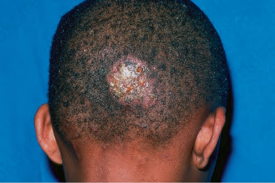
Fig. 18.264 Kerion: in addition to alopecia there is marked erythema and matting of hairs due to purulent exudate. By courtesy of R.A. Marsden, MD, St George’s Hospital, London, UK.
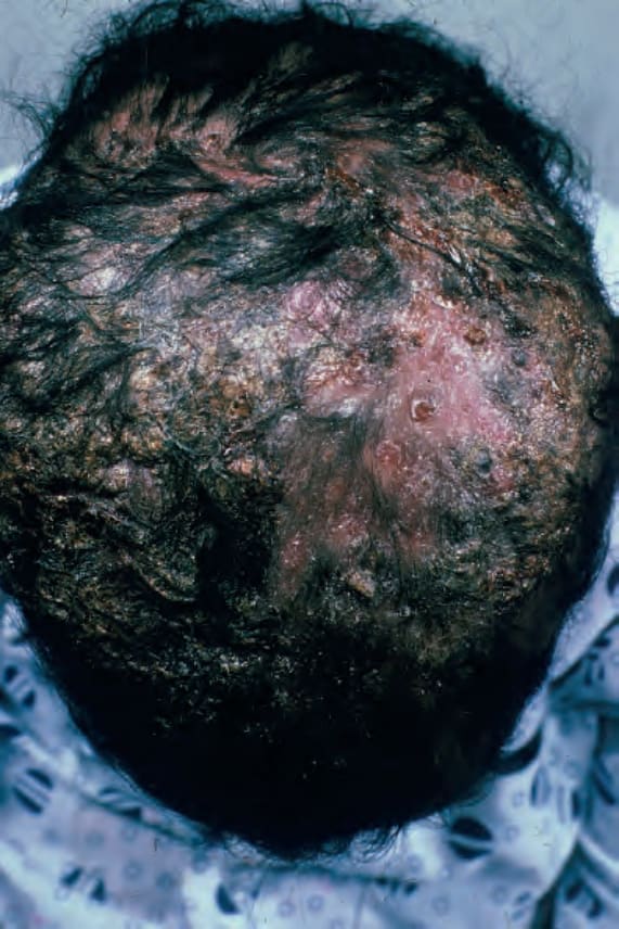
Fig. 18.265 Kerion: there is marked alopecia and crusting. From the collection of the late N.P. Smith, MD, the Institute of Dermatology, London, UK.
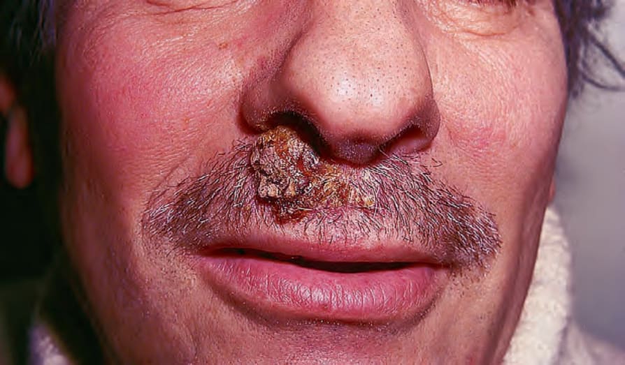
Fig. 18.266 Kerion: in males, dermatophyte infection of the beard area may also present as a kerion. By courtesy of R.A. Marsden, MD, St George’s Hospital, London, UK.
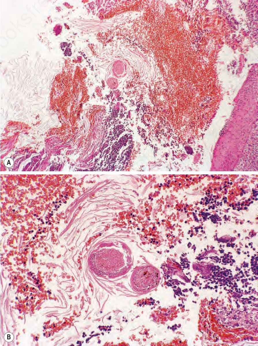
Fig. 18.268 Kerion: (A) crust scrapings from a patient with a typical scalp lesion. In addition to blood, keratinous debris and numerous neutrophils, two hair shafts are present in the center of the field. (B) High-power view showing numerous fungal spores coating the outside of the shaft.
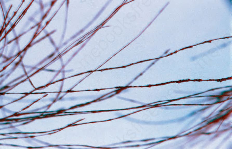
Fig. 18.269 Favus: scarring alopecia due to infection by Trichophyton schoenleinii. By courtesy of R.A. Marsden, MD, St George’s Hospital, London, UK.