Tinea capitis
Tinea capitis
Dermatophyte infections of the scalp are characterized by involvement of the hair shaft by pathogenic fungi. The pattern of hair invasion, related to the type of dermatophyte, determines the degree and site of hair damage and the clinical picture. Patients therefore present with variable features including hair loss with scaling, follicular inflammation, pustulation, and kerion formation, often in association with drainage lymphadenopathy. A carrier state is recognized. Infection depends on contact with spores and follicular trauma. Tinea capitis (scalp ringworm) frequently presents as small epidemics (e.g., in schools). Disease may develop as a consequence of sharing combs or hairbrushes. Tinea capitis is the most common dermatophytosis of childhood. The disease may also occur in adults, the elderly, and even infants. The past two decades have seen a rise in its prevalence and a change in the pattern of etiological agents, both in Europe and the United States.
Infections caused by Microsporum canis and Microsporum audouinii
Clinical features
M. canis and M. audouinii grow on the outside of the hair shaft, an ectothrix type of hair involvement.1–3 The lesions present as areas of alopecia, with numerous broken-off, dull hairs (Fig. 18.262). Some scaling is present, but overt inflammation is not marked. The infected areas are recognized by fluorescence under Wood lamp (Fig. 18.263). The process commonly affects children, boys much more often than girls. M. ferrugineum may also cause this picture.
M. canis and M. audouinii were once the most common causes of tinea capitis in North America and Western Europe.3,4 M. canis, a zoophilic dermatophyte, is the organism most frequently implicated in cases of tinea capitis in Europe. Urban areas of Europe (and France in particular) have shown a shift toward infection with M. audouinii, an anthropophilic dermatophyte.5,6
Histologic features The fungal arthrospores coat the outside of the hair shaft and the hyphae extend into the hair shaft down to the level of the mid follicle. The epidermis shows some acanthosis and patchy parakeratosis, and there is usually a
mixed inflammatory infiltrate in the superficial dermis, more marked with M. canis.
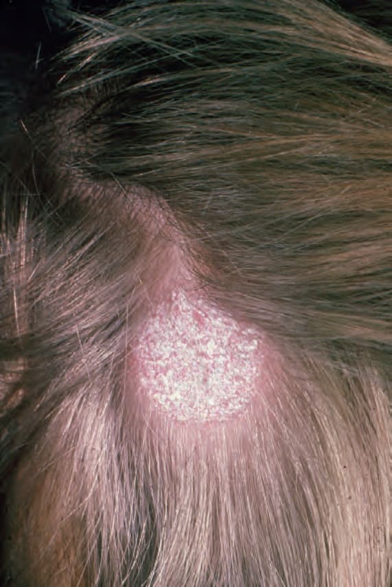
Fig. 18.262 Tinea capitis: there is marked hair loss. In this example scaling and crusting are pronounced. From the collection of the late N.P. Smith, MD, the Institute of Dermatology, London, UK.
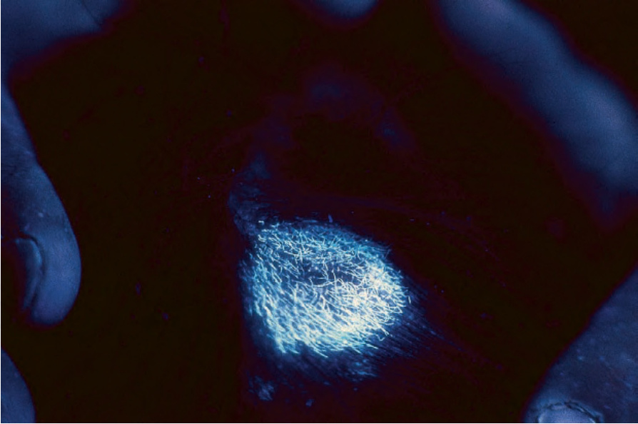
Fig. 18.263 Tinea capitis: note the characteristic fluorescence under Wood light. By courtesy of M.M. Black, MD, Institute of Dermatology, London, UK.