FUNGAL INFECTIONS
FUNGAL INFECTIONS
Fungal infections include:
• superficial variants involving skin, hair, nails, and mucous membranes, for example, ringworm (dermatophytosis), and the dermatomycoses (tinea versicolor and candidiasis),
• subcutaneous lesions,
• disseminated infection.1,2
Ringworm fungi include three species: Microsporum, Trichophyton, and Epidermophyton.3,4 Epidermophyton invades epidermal keratin while Microsporum and Trichophyton also affect the hair. Cutaneous ringworm on nonhairy skin presents as slowly enlarging, scaly, erythematous, annular lesions with central clearing (Fig. 18.261).
Dermatophytes can infect the keratin of the stratum corneum, hair, or nail, without extending into deeper parts of the skin. They may also be associated with intradermal spread (Majocchi granuloma). Causative organisms may be anthropophilic, zoophilic, or geophilic. With few exceptions, identification of pathogenic fungi is better served by culture rather than by histologic scrutiny.5–7 Dermatophytes use the soluble nonkeratin parts for nutrition and rely on the keratin for protection from serum and the host response.8,9 The keratin is penetrated by means of putative keratinases. Other virulence factors include elastase and proteinases.9,10 T. rubrum produces mannan, which suppresses or diminishes the host immune response, presumably by inhibiting critical steps in antigen presentation or
925 Tinea capitis
processing.9,11 The epidemiology and pathogenesis of dermatophytosis is complex and beyond the remit of this text. For suitable review articles, the reader is referred to references 1, 3, 4, and 10.
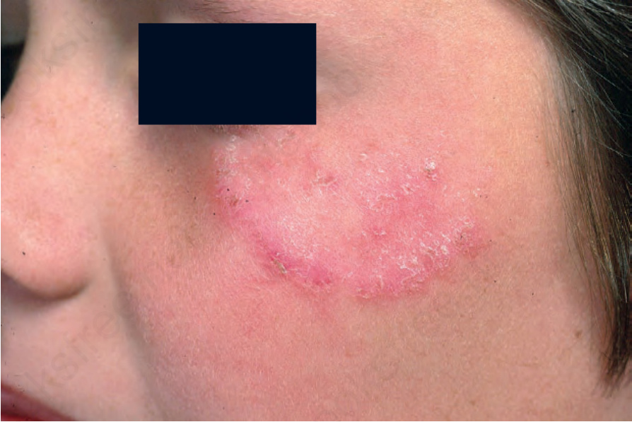
Fig. 18.261 Tinea corporis: note the annular configuration and erythematous margin. From the collection of the late N.P. Smith, MD, the Institute of Dermatology, London, UK.