Cutaneous diphtheria
Cutaneous diphtheria
Clinical features Diphtheria is a highly contagious, vaccine-preventable, usually upper respiratory tract infection caused by the Gram-positive bacillus Corynebacterium diphtheriae. An asymptomatic carrier state also exists.1,2 Cutaneous diphtheria is an uncommon condition traditionally ascribed to C. diphtheriae.3,4 Recent years, however, have seen an increasing role for C. ulcerans in the etiology of cutaneous diphtheria, especially in European countries.1,5–7 Toxogenic and nontoxogenic strains of both organisms exist.6–18 Rare cases of C. pseudodiphtheriticum and C. pseudotuberculosis infection have also been reported.1,19 Although cutaneous diphtheria is essentially a tropical condition, increasing tourism to tropical regions and a decline in adult booster vaccination against diphtheria have resulted in numerous cases being reported from developed countries, especially among returning travelers who have visited regions where the infection is endemic.1,2,4,8–11,14,17,20–23 The disease usually results from inoculation of organisms into pre-existing lesions such as burns, ulcers, abrasions, and eczematous rashes, and has even occurred following tattooing; cutaneous diphtheria may also manifest in apparently normal skin.3,4,7,8,20 Cutaneous C. diphtheriae infection has been reported among intravenous drug users and homeless individuals, especially in impoverished urban areas.16,24 Contact with infected domestic animals or occupational exposure to agricultural animals are additional risk factors for C. ulcerans infection.6,12,18
The lower legs and feet are sites of predilection, but sites such as the face, trunk, hands, and even the genitalia may be involved.3,6,7,9,10,15–17,25 Initially, there is a vesicle or pustule. This later evolves into an ulcer which is often reddish-purple in color, with rolled and undermined borders, and a yellow-gray membrane or dark crust covering its base.3,20 The ulcers are painful initially but are later hypoanesthetic.3 Regional lymphadenopathy may occur, and toxicogenic strains may result in systemic complications involving the nervous system or heart.1–3
Histologic features Histologic examination of the ulcer reveals a necrotic epidermis and dermis. The dermal base of the ulcer contains necrotic debris, fibrin, and an admixture of acute and chronic inflammatory cells.2 Since the club-shaped and beaded Gram-positive rods are often difficult to visualize in histologic material, microbiological examination of swabs from the center of the lesion is required for confirmation of the diagnosis.3 Co-infection with other bacterial organisms, however, is a frequent occurrence.16,17
may occur concurrently with erythrasma and trichobacteriosis, resulting in the so-called corynebacterial triad.3
Pathogenesis and histologic features The pits are the consequence of dissolution of the stratum corneum induced by the pathogenic bacteria. In vitro studies have shown that both D. congolensis and K. sedentarius produce keratinolytic enzymes, thus accounting for the superficial defects in the stratum corneum.8,17
The early lesions demonstrate stratum corneum pallor. Biopsies of the plantar pits show small defects in the upper stratum corneum, the walls
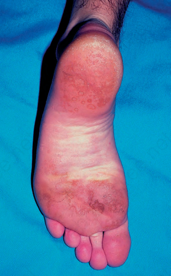
Fig. 18.238 Pitted keratolysis: note the tiny pits on the weight-bearing aspect of the foot. By courtesy of S. Glassman, MD, Division of Dermatology, University of Witwatersrand, Johannesburg, South Africa.