Trichobacteriosis
Trichobacteriosis
Clinical features Trichobacteriosis is the preferred and more contemporary designation for trichomycosis, a bacterial infection caused by different species of corynebacteria. Although traditionally ascribed to Corynebacterium tenuis, recent advances in molecular taxonomy have shown that the usual etiological agent belongs to the CDC-G/LD (or so-called LD2) group, which corresponds to C. flavescens.1–3 The Gram-positive bacteria invade the cuticle of the hair. Although it was initially suggested that they adhere to the hair shaft by producing a cement-like substance, this view was subsequently challenged and it was later proposed that the material that provides support for the organisms is the apocrine sweat.4–6 Disturbances in apocrine/apo-eccrine sweat production and associated bacterial proliferation appear to play a crucial pathogenetic role.1,3 The disease typically involves the axillary hair (trichobacteriosis/trichomycosis axillaris) and exceptionally may be seen in the pubic hair and scrotum (trichobacteriosis/trichomycosis pubis) (Fig. 18.236.7–11 Trichobacteriosis is characterized by yellow, red, or black nodules along the hair shaft. These nodules may be confused with nits. However, the nodules in trichobacteriosis fluoresce under Wood light. In black piedra, the nodules are usually black and the disease mainly involves the scalp. There have nevertheless been single case reports of scalp involvement in infants (trichobacteriosis capitis).12,13 Distinction from white piedra may be more difficult as the disease has a wide anatomical distribution and it has even been suggested that the latter disease is the result of a synergistic interaction between corynebacteria and Trichosporon beigelii, the organism
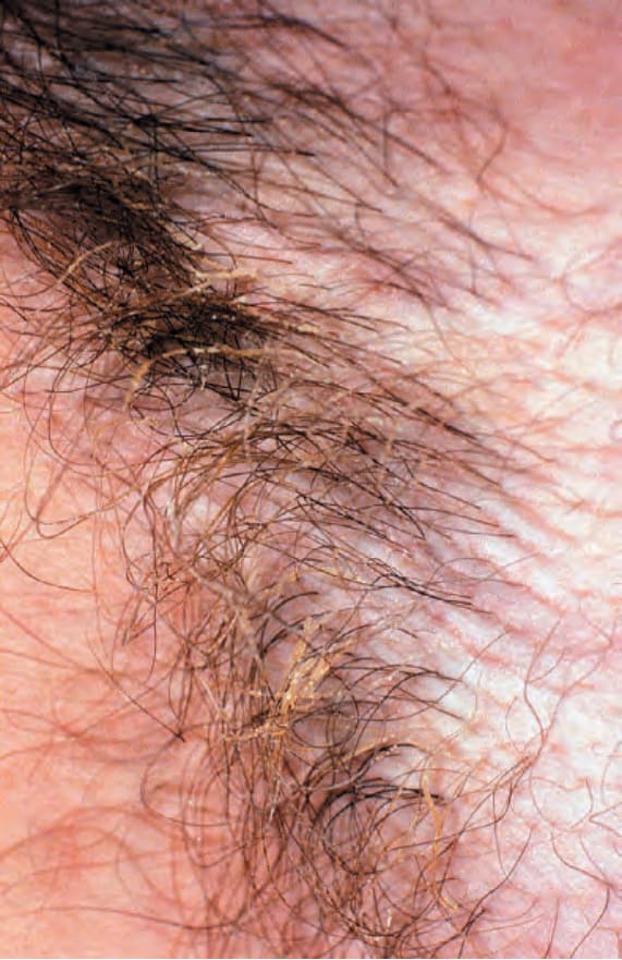
Fig. 18.236 Trichomycosis: this matted appearance of the hair results from the presence of multiple tiny nodules. By courtesy of the Institute of Dermatology, London, UK.