Erythrasma
Erythrasma
Clinical features Erythrasma is a bacterial infection caused by Corynebacterium minutissimum, a Gram-positive bacillus.1–3 It characteristically presents as asymptomatic, well-defined, scaly red patches on the inguinal and intergluteal skin (Fig. 18.234). It has a predilection for obese and diabetic patients and it is more common in areas with a humid and hot climate. The clinical diagnosis is easy because of the demonstration of a typical coral-red fluorescence under Wood light.4,5 This fluorescence is the result of production of coproporphyrin III by the organism. In exceptional cases, fluorescence is not seen.6 Erythrasma rarely presents as a disciform eruption with an atrophic appearance involving nonintertriginous areas.7,8 Involvement of the feet (particularly the toe webs) and toenails has also been documented.9–13 Rarely, lesions on the feet have a vesiculobullous appearance.14 Nail involvement is
Skin involvement in the form of cutaneous hyperpigmentation is relatively common, occurring in 17% to 40% of patients.5,11,12 Rarely, erythema nodosum-like subcutaneous nodules may develop as a result of infective panniculitis. The latter comprise painful red-brown nodules. Lesions may occur on the thighs, arms, buttocks, lower legs, and, rarely, the chin and neck.10,13–16 Erythema nodosum proper is another potential manifestation; it may also be encountered in those who develop IRIS.8,13 The evolution of ENL-like lesions as a manifestation of IRIS in treated Whipple disease has also been documented.9
Pathogenesis and histologic features Although T. whipplei is a ubiquitous organism in the environment, Whipple disease remains a very rare condition, with an estimated prevalence of 1.1 per 1 million population and an annual incidence of around 6 per 10 million population.1,17 Susceptible individuals have defects in T-cell immunity and impaired ability of macrophages to degrade the intracellular organisms.1,3,18
The subcutaneous nodules have the appearance of a predominantly septal panniculitis, with a conspicuous infiltrate of foamy macrophages. Careful examination reveals cytoplasmic distension of these histiocytes by PAS-positive, diastase-resistant intracytoplasmic material representing degenerate bacteria.9,10,15,16 These macrophages closely resemble those encountered in duodenal biopsy material. The viable bacilli have a characteristic double-walled (trilaminar) appearance on ultrastructural examination.1,13 In rare instances, granulomas may be observed in the dermal compartment overlying the septal panniculitis.10 Dermal lymphangiectasia has also been described.9 The organism may be cultured in specialized laboratories. PCR and immunohistochemical methods for confirmation of the diagnosis exist.10,11,17–20
A
Differential diagnosis Whipple disease should be distinguished from histoplasmosis, histoid LL, and infection with MAI complex, as all of the aforementioned conditions are characterized by intracytoplasmic organisms contained within macrophages. PAS-positive fungal yeasts, however, are observed in histoplasmosis, whereas Ziehl-Neelsen and Wade-Fite stains will confirm the presence of acid-fast mycobacterial bacilli in MAI infection and LL, respectively.
The term ‘pseudo-Whipple disease’ was used to describe two recently reported cases who presented with clinical and histologic features suspect for cutaneous/subcutaneous Whipple disease, but in whom no T. whipplei organisms could be detected by PCR. The lesions resolved on treatment with penicillin; a bacterial (staphylococcal) infection, however, could only be proven in one of the patients.21
In those exceptional cases of cutaneous Whipple disease demonstrating intradermal granuloma formation, the condition should be distinguished from other causes of granulomatous dermatitis.10
B
912 Infectious diseases of the skin
characterized by hyperkeratosis and onycholysis. Erythrasma may coexist with a dermatophyte infection or, rarely, pityriasis versicolor.15,16 An association with trichomycosis (trichobacteriosis) axillaris and pitted keratolysis may also occur; this has been referred to as the corynebacterial triad.5,17,18 A biopsy is only exceptionally performed as the diagnosis is confirmed by the use of Wood light or scrapings stained by Gram, PAS, Grocott, Giemsa, or methylene blue.
C. minutissimum has rarely been associated with bacteremia, abscess formation, cellulitis or visceral involvement in immunocompetent or immunocompromised patients.3,19–22
Histologic features A skin biopsy usually appears unremarkable when stained with H&E except for mild hypergranulosis and occasional superficial perivascular lymphocytes. The special stains mentioned before, particularly Gram, show the presence of bacilli in the stratum corneum (Fig. 18.235).
previously implicated in white piedra.14 Dermoscopy is a potentially useful diagnostic adjunct.13,15,16 Trichobacteriosis has been reported in association with erythrasma and pitted keratolysis.3,17,18
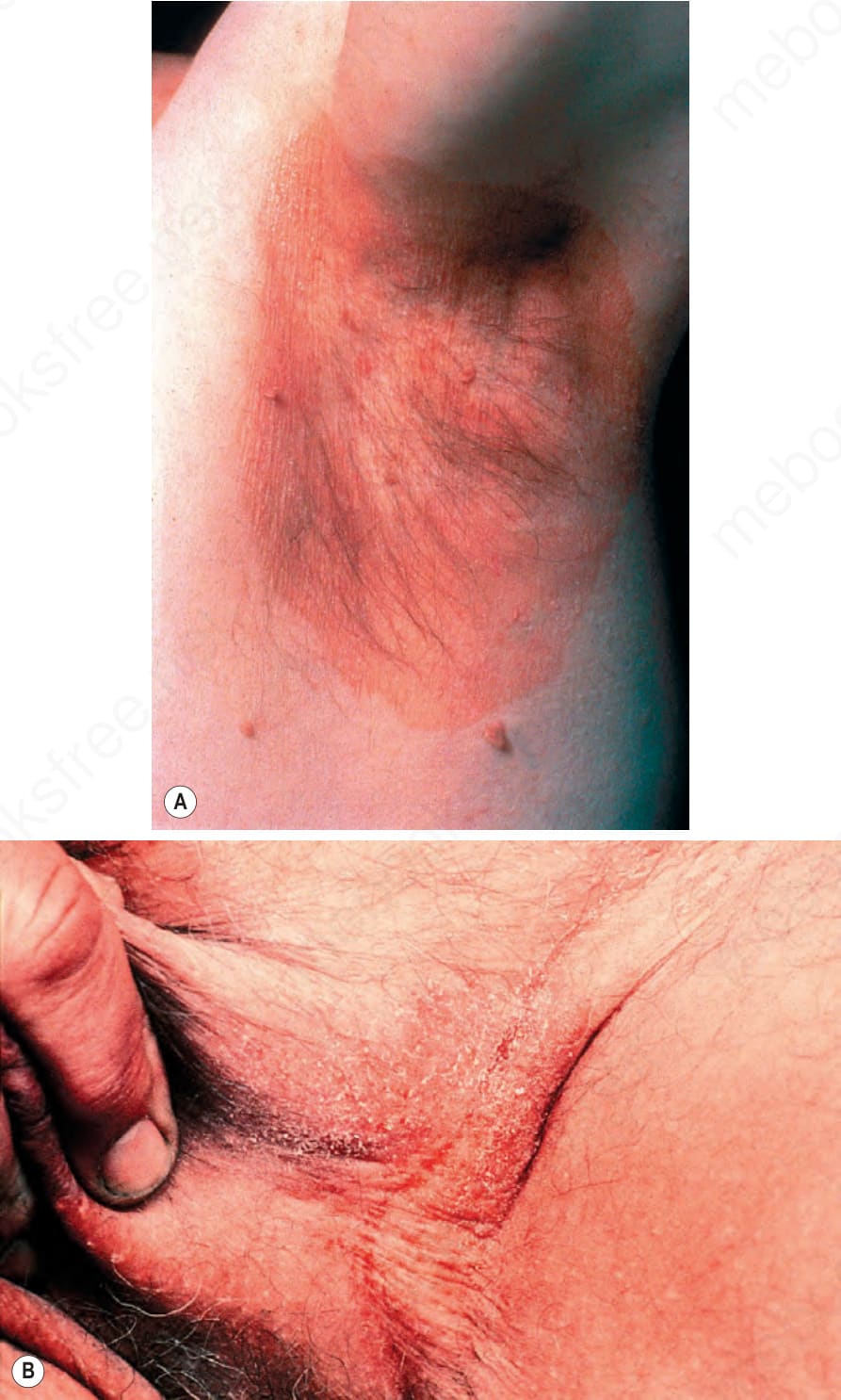
Fig. 18.234 Erythrasma: (A) note the well-demarcated axillary scaly red patch; (B) there is a scaly inguinal patch with associated hyperpigmentation. By courtesy of the Institute of Dermatology, London, UK.
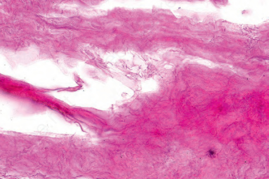
Fig. 18.235 Erythrasma: bacilli are evident in the stratum corneum.
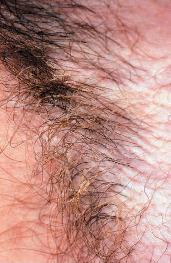
Fig. 18.236 Trichomycosis: this matted appearance of the hair results from the presence of multiple tiny nodules. By courtesy of the Institute of Dermatology, London, UK.