Nocardiosis
Nocardiosis
Clinical features Nocardia is found in soil and rotting vegetation worldwide and man is only rarely infected.1–4 Initially, three main pathogenic species were recognized:
• Nocardia asteroides, which is most common in North America,
• Nocardia brasiliensis in South America,
• Nocardia caviae in Southeast Asia.1,3,5
906 Infectious diseases of the skin
Other species of Nocardia have since been associated with infection in humans, including N. transvalensis, N. otididiscaviarum, N. nova, N. farcinica, N. paucivorans, N. abscessus, N. cyriacigeorgici, N. asiatica, N. vinacea, N. beijingensis, N. araoensis, and N. neocaledoniensis.1,4,6–23 More than 50 species of Nocardia are now known to exist.6 Infection complicates inhalation or direct inoculation into a wound.1,3,6 Nocardiosis therefore represents a respiratory illness, with or without dissemination (the majority), or a primary cutaneous disease.1,2,6,24
N. asteroides, N. caviae, and N. farcinica most often affect immunocompromised hosts, causing pulmonary lesions from which systemic dissemination may involve the skin.3,25 Predisposing factors include steroid therapy, HIV infection, and solid organ transplantation.1,4,12–14,26–29 CNS involvement is an important cause of morbidity and high mortality.12,27 Primary cutaneous infection has been reported in an immunocompetent adult following a cat scratch.30 Dissemination is a frequent complication of N. paucivorans infection in both immunocompromised and immunocompetent hosts.15 Nocardia infections have been recorded among patients who have received immunomodulatory therapy for inflammatory bowel disease.31
A
N. brasiliensis causes primary skin lesions in immunocompetent individuals and can also cause primary pulmonary lesions.4,32 The cutaneous lesions are varied and usually follow trauma. They include a mycetoma, usually on the limbs, and a sporotrichosis-like pattern (including a cervicofacial variant that occurs in children), i.e., with multiple lesions following the line of lymphatics.3,33–40 In addition, superficial nodules, ulcers, and abscesses, with or without fistulae and pustules, may occur (Fig. 18.222).41–44 Some more trivial infections resemble staphylococcal infections and are usually self-limiting; the deeper infections are progressive and can be destructive without treatment. Dissemination of primary cutaneous nocardiasis is exceptionally uncommon.45 Infection has been reported after an insect bite.46 Lymphocutaneous nocardiosis may also be caused by N. transvalensis and N. araoensis.7,22 Mycetomas due to N. nova and N. otididiscaviarum have been reported.8,11 N. otididiscaviarum is a rare cause of cellulitis.10 Nocardia spp. are a rare infectious cause of neutrophilic eccrine hidradenitis.47
B
Pathogenesis and histologic features Nocardia is a Gram-positive, partially acid-fast aerobic beaded rod, which grows with branching filaments in a way similar to Actinomyces (Fig. 18.223).48 Infection by N. asteroides, a relatively avirulent organism, is usually by inhalation. N. brasiliensis is more virulent and can infect the immunocompetent through inoculation of soil into skin. It is the most commonly identified organism in the sporotrichoid form of nocardiosis.4,34,36,38,39
Grains, analogous to the sulfur granules of actinomycosis, can develop in the mycetoma lesions of the immunocompetent. Filamentous growth is
907 Botryomycosis
A
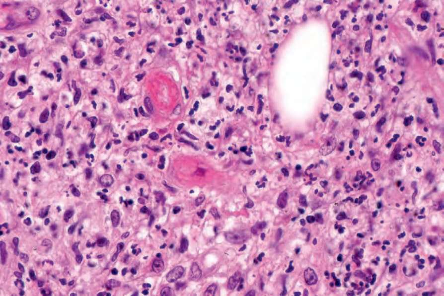
Fig. 18.219 Erythema nodosum leprosum: several small vessels show fibrinoid necrosis. This type II reaction develops on the basis of an immune complex-mediated vasculitis.

Fig. 18.220 (A, B) Rhinoscleroma: in addition to lymphocytes and numerous plasma cells, foamy macrophages (Mikulicz cells) are present.
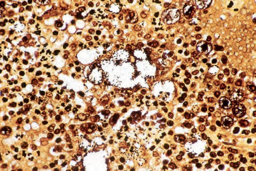
Fig. 18.221 Rhinoscleroma: numerous organisms are revealed by the Warthin-Starry reaction. By courtesy of S. Lucas, MD, Institute of Dermatology, London, UK.
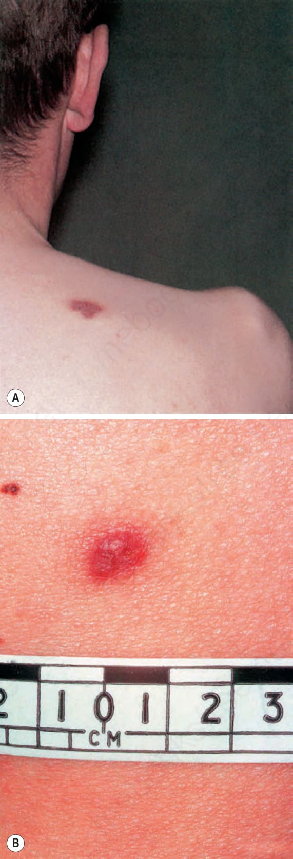
Fig. 18.222 Nocardiosis: (A) this cutaneous nodule developed in an immunocompromised young male; (B) a different lesion is shown in close-up. By courtesy of R.A. Marsden, St George’s Hospital, London, UK.
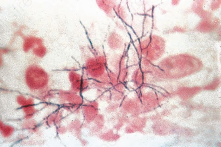
Fig. 18.223 Nocardia: the organisms appear mainly as irregularly staining filaments in this specimen, but a variety of forms, including rods and cocci, is often seen. By courtesy of A.E. Prevost, MD, and H.P. Lambert, MD, St George’s Hospital, London, UK.