Brucellosis
Brucellosis
Clinical features Brucellosis is a zoonotic infection by Brucella spp. such as B. melitensis, B. abortus, B. canis, and B. suis.1–3 The organism is a Gram-negative bacillus, and infection is acquired either by ingesting contaminated, unpasteurized milk/milk products, or by handling infected animal products (contact brucellosis). Human-to-human transmission does not occur. Brucellosis results in either an acute febrile illness or a chronic systemic disease characterized by fever, malaise, sweats, arthralgia, myalgia, and/or hepatosplenomegaly.1–3 Although the disease is endemic in several countries bordering the Mediterranean Sea, the majority of reports of human infection appear to have come
872 Infectious diseases of the skin
Histologic features
The histologic features vary according to the type of cutaneous lesion biopsied. Biopsies obtained from the erythematous papular and maculopapular lesions show a perivascular and periadnexal infiltrate of lymphocytes and histiocytes, sometimes accompanied by epithelioid histiocytes and multinucleated giant cells.7,11 Erythrocyte extravasation is sometimes seen, and inflammatory cells may infiltrate the overlying epidermis.10 Erythema nodosum-like nodules are characterized by a perivascular lymphohistiocytic infiltrate centered on the deep dermis and superficial subcutis.7,12 There is associated vascular endothelial swelling and luminal thrombosis, sometimes with foci of necrosis. Accompanying granulomatous inflammation is not uncommon.10 Erythema multiforme lesions and leukocytoclastic vasculitic lesions exhibit the usual histologic changes associated with these conditions.
measure 1–5 mm or more in diameter and may sometimes resemble an insect bite.1
Other rare cutaneous manifestations include a nonpruritic macular or maculopapular rash, erythema nodosum, urticaria, erythema marginatum, erythema annulare, and thrombocytopenic purpura. A case with cutaneous lesions resembling those of Sweet syndrome has been reported.11 Fever and malaise are occasional symptoms. Less common additional features include headache, nausea, vomiting, arthralgia, and splenomegaly.
Patients invariably develop lymphadenopathy in the drainage region, usually within 1–3 weeks of the initial lesion. The enlarged nodes are tender and often persistent, with lymphadenopathy lasting up to 2 months or more. Suppuration is not uncommon. Conjunctival or eyelid lesions may be associated with preauricular lymphadenopathy (Parinaud syndrome).12 The only consistently abnormal laboratory function test is a moderately raised erythrocyte sedimentation rate. Purported cases of disseminated cat scratch disease occurring as a manifestation of AIDS probably represent examples of advanced BA (see below).13,14
Brucella organisms are rarely visualized in histologic material. The diagnosis is therefore confirmed by culture, serology or, PCR.1,3,7,10,23
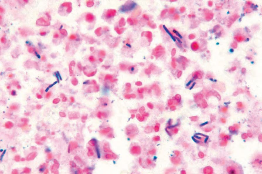
Fig. 18.125 Anthrax: numerous elongated Gram-positive bacilli are present. Reproduced with permission from Mallon E, McKee PH. Extraordinary case report: cutaneous anthrax. American Journal of Dermatopathology. 1997; 19: 79–82.
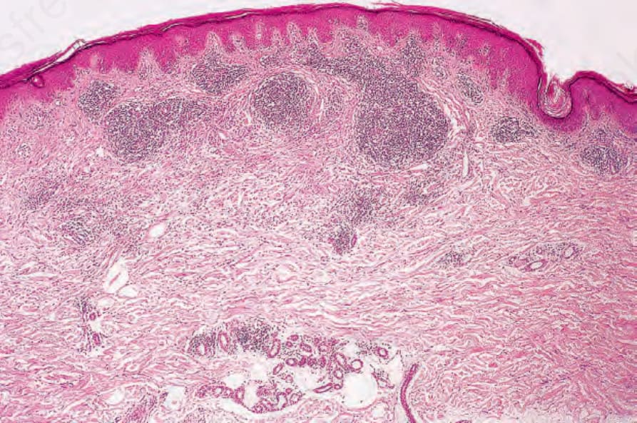
Fig. 18.126 Cat scratch disease: an irregular ill-defined focus of necrobiosis is surrounded by a nodular lymphocytic infiltrate.