Meningococcal septicemia
Meningococcal septicemia
Clinical features Meningococcal septicemia (meningococcemia) is due to the Gram-positive diplococcus Neisseria meningitidis.1–6 In its acute form, this is a very serious condition with a high mortality, which affects children in seasonal epidemics. The infection may occasionally be encountered in adults.6 Occurrence in the neonatal period, however, is rare.7 The organism is spread via droplet inhalation from upper respiratory tract infections. Meningitis is the most frequently encountered manifestation of meningococcal infection. Children with acute meningococcal septicemia develop widespread purpura that shows a predilection for the trunk and limbs. Ecchymoses may also be a feature. In the more chronic variant (chronic meningococcemia), patients present with vasculitis-like lesions, particularly nodules, and palpable purpura.8
Pathogenesis and histologic features The histologic features are essentially those of a leukocytoclastic vasculitis.9 Superimposed DIC may also be present. The hemorrhagic skin lesions and vascular thromboses are attributable to up-regulation of tissue factor leading to coagulation, and by inhibition of fibrinolysis by plasminogen activator inhibitor.10 Impairment of the protein C anticoagulation pathway also plays an important role.11 Experimental evidence has revealed that adhesion of the organism to the dermal microvascular endothelium leads to local vascular damage, thrombosis, and purpura.12 Capsular polysaccharides and lipooligosaccharides play key roles in the evasion of killing by host complement.13
have been documented.10–12 Lobular capillary hemangioma (pyogenic granuloma)-like lesions of the penile shaft have also been described.13 Primary digital gonorrhea has been recorded.10
Pathogenesis and histologic features Disseminated lesions show variable epidermal changes ranging from edema accompanied by a neutrophil infiltrate with purpura, to vesiculation, pustulation, and eventually necrosis.6 In the dermis, the histologic features are essentially those of a neutrophil-mediated acute vasculitis, often accompanied by thrombosis.14 Very occasionally, Gram-negative diplococci may be identified.
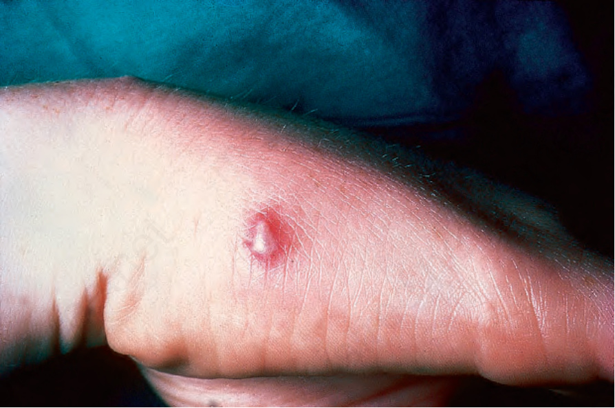
Fig. 18.118 Gonococcemia: pustules are commonly found on the hands and feet. By courtesy of R.N. Thin, MD, St Thomas’ Hospital, London, UK.