Milker nodule
Milker nodule
Clinical features Milker nodule (or paravaccinia) is caused by a parapox virus, distinct from that which causes cowpox.1,2 It occurs as a localized lesion on the udders of cows and causes little systemic disturbance. It may be recurrent in the same herd. It is usually acquired by man by inoculation, but since the virus is viable in a dried state, indirect fomite infection is possible. Small outbreaks involving several patients have been recorded.2 The incubation period is around 5 days; some two to five red papules then develop, which gradually become bluish tender nodules.2 The overlying epidermis is at first tense and shiny, but becomes opaque and gray (Fig. 18.68). The center of the lesion is crusted and slightly depressed. The surrounding skin often shows lymphangitis, but despite this the lesion has the appearance of a tumor and is well circumscribed. There are few systemic symptoms, but there may be an associated short-lived papulovesicular eruption on the upper limbs and occasionally on the legs. The main nodular lesions resolve, without scarring, in 4–6 weeks.
Histologic features Milker nodule virus measures 160–260 nm and is ellipsoid in shape.2 It is characterized by spirally arranged tubules. The histologic features are indistinguishable from those seen in orf infection (see below).3,4 A CD30-positive atypical dermal lymphoid infiltrate may sometimes be encountered, and confusion with a CD30-positive lymphoproliferative disease is possible.5 In the latter, the CD30-positive cells are usually in clumps while in Milker nodule the CD30-positive cells are scattered between other inflammatory cells. This is, however, not entirely consistent, and in some cases clusters of CD30-positive cells may be seen.
pasture is dry and results in minor abrasions of the labial mucosa of the sheep. Transmission to man, most often males and usually sheep-handlers, is usually by direct inoculation from infected lesions, but it may also result from contact with contaminated objects such as fences and shears.5 Orf may also be seen in goats.3–5,7 In one English study, 23% of individuals employed or living on a sheep farm reported having had the condition.8 Nonoccupational acquisition of the infection has been described following a sacrificial feast.9
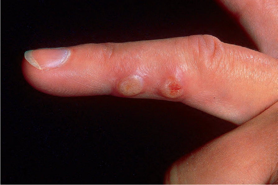
Fig. 18.68 Milker nodule: the blister roof has an opaque appearance and there is surrounding erythema. By courtesy of the Institute of Dermatology, London, UK.
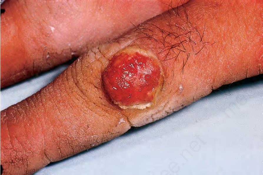
Fig. 18.70 Orf: in this example there is a markedly hemorrhagic component. By courtesy of M.M. Black, MD, Institute of Dermatology, London, UK.