Myiasis
Myiasis
Clinical features The term ‘myiasis’ is derived from the Greek word for fly (myia). The condition is the result of cutaneous infestation with maggots (larvae) of fly species from the order Diptera. Infestation with larvae of the human botfly,
Three clinical forms may occur in human skin:
• furuncular myiasis, with boil-like lesions occurring on exposed parts of the body such as the scalp, face, or limbs if botfly associated, and usually on nonexposed sites such as the buttock, thighs, breasts, or trunk if tumbu fly associated;
• wound myiasis due to deposition of larvae of flies such as Cochliomyia americana, Chysomia bezziana, or Lucilia sericata in pre-existing wounds;
• migratory (creeping) myiasis, a zoonosis caused by larvae of Hypoderma bovis or Gasterophilus intestinalis.1,2,5–7
Furuncular myiasis presents initially as an erythematous papule which enlarges to form a 1–3 cm painful, sometimes crusted boggy plaque or ‘boil’ with a central punctum through which serosanguinous fluid may discharge (Fig. 18.389). A mature 15–20 mm larva eventually emerges within 5–10 weeks.1,2,4
969 Onchocerciasis
Histologic features The cross section through the larva is distinctive in its appearance. Depending on the plane of sectioning, polarizable structures such as the thick cuticle covered with spines, the two curved mouth hooks and/or the respiratory spiracles may be observed in histologic material (Fig. 18.390). Biopsies which include the surrounding skin may show an ulcerated epidermis, a cavity lined by acute and chronic inflammatory cells, and a neighboring dermal inflammatory infiltrate comprising lymphocytes, plasma cells, eosinophils, giant cells, and Langerhans cells.1–3,8,9
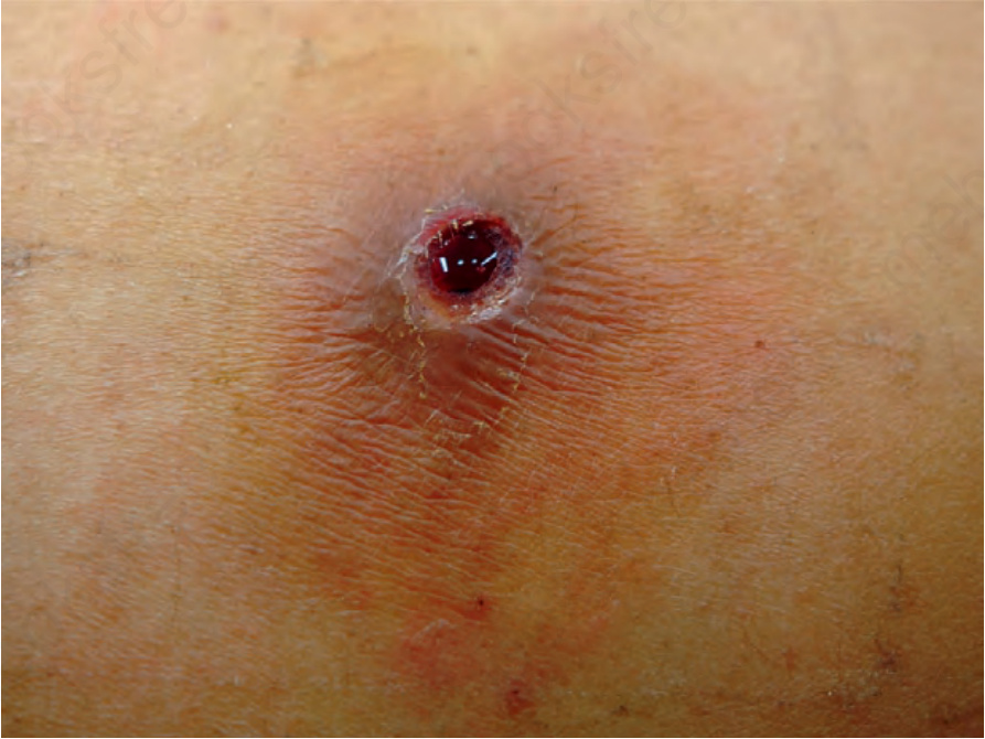
Fig. 18.389 Myiasis: characteristic furuncular lesion in a patient with botfly infestation. By courtesy of F. Bravo, MD, Lima, Peru.
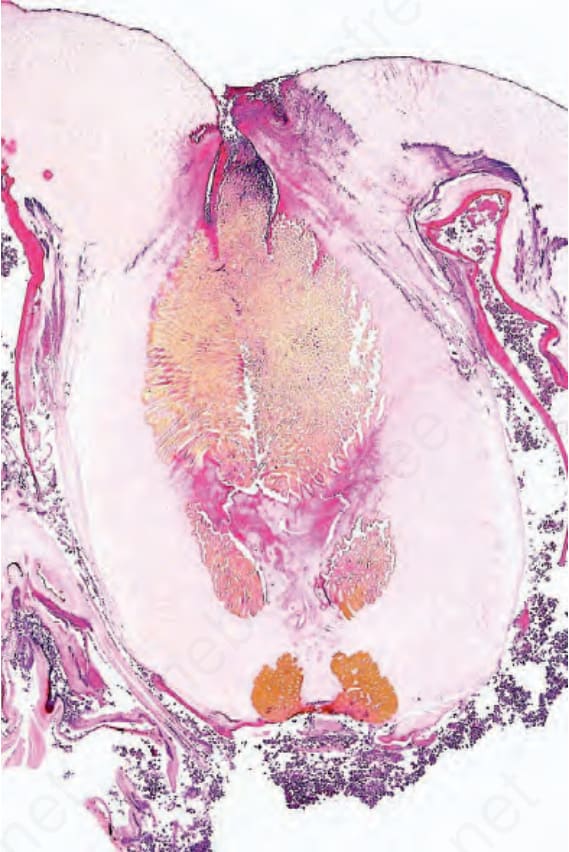
Fig. 18.390 Myiasis: intradermal botfly larva showing characteristic respiratory spiracles.