Bowenoid papulosis
Bowenoid papulosis
Clinical features Bowenoid papulosis (koilocytosis with intraepithelial neoplasia) is a clinicopathological entity that bears marked histologic similarity to koilocytosis, SIL/CIN, and Bowen disease. Although the term is no longer used by the International Society for Study of Vulval Disease (ISSVD) and some have questioned the validity thereof, many clinicians believe that it represents a distinctive clinicopathological entity, and we have decided to describe it in this chapter. Clinically, it is quite different from genital Bowen disease in that multiple small papules develop over a short time scale in young people. Prognosis is uncertain; many patients do not show evidence of progression, but a small proportion may develop invasive tumor and, on occasion, this may have metastatic potential. It is usually associated with HPV16 or 18, but occasionally HPV types 31–35, 39, 42, 49, and 51–54 are detected.1–10 Although uncommon, some cases may be associated with mixed infection by different HPV types.10,11 A unique HIV-associated case with genital and extragenital (lip) lesions caused by two separate HPV types (HPV16 and HPV32, respectively) has been described.12 E6 and E7 viral oncoproteins of high-risk HPV types induce overexpression of p16 and human telomerase reverse transcriptase.13
835 Epidermodysplasia verruciformis
Bowenoid papulosis most often presents as multiple reddish-brown, sometimes lichenoid, discrete papules, but occasionally these become a confluent plaque. Papules, on average 4 mm in diameter, are found on the penis, vulva, perianal region, and perineum. Extragenital sites of occurrence include the face, neck, and fingers.14–16 The lesions are sometimes pigmented.2 A case of oral bowenoid papulosis in an HIV-infected male has been reported.17 Bowenoid papulosis manifests in young, sexually active adults in contrast to true Bowen disease, which occurs in an older age group. Genital Bowen disease is, however, also often associated with HPV16.18 The occurrence in childhood should raise suspicion of sexual abuse.7 Genital bowenoid papulosis has been associated with periungual bowenoid dysplasia.19 Bowenoid papulosis with concurrent Bowen disease has been reported in a patient with systemic lupus erythematosus (SLE).20
Spontaneous regression is uncommon.21 As progression to frank invasive carcinoma in bowenoid papulosis is rare, these lesions are best managed conservatively. However, bowenoid papulosis may be resistant to treatment and may be characterized by a prolonged course in immunosuppressed patients.6 Bowenoid papulosis has also been associated with oral warts and lingual carcinoma.8 Patients with bowenoid papulosis and HPV infection may be primarily immunosuppressed due to diminished T-helper (Th) cell levels (non-HIV-associated).6,18 The condition may also occur in organ transplant recipients.22 Penile bowenoid papulosis is associated with a high risk of the consort developing cervical dysplasia.23,24 Consequently, female patients and consorts should regularly have cervical smears.
A
Histologic features A bowenoid papulosis lesion consists of a well-circumscribed area of acanthosis producing a raised plaque or dome, which is hyperkeratotic and sometimes shows superficial epithelial vacuolation.25,26 The keratinocytes may show nuclear hyperchromatism and pleomorphism. There is variable dyskeratosis.
B
especially squamous cell carcinomas.1–5 The lesions of affected individuals are the result of infection with a wide range of HPV subtypes including 3, 5, 8–10, 12, 14, 15, 17, 19–25, 28, 29, 36–38, 46, 47, 49, 50, 51, and 59.1,6–9 The vast majority of these are β-HPV genotypes.10 The more common flat warts, caused by HPV3 and HPV10, may also occur in these patients but have an extensive distribution pattern; they may form plaques and can be persistent.11 These are seen most often on the arms, legs, face, and the dorsum of the hands (Figs 18.27–18.29).7 The specific EV subtypes of HPV cause reddish, or pigmented or depigmented, scaly flat macular plane warts, mainly on the trunk, but also on the face, neck, and arms.2 Clinically, they resemble pityriasis versicolor (Fig. 18.30). Some patients, especially those who are dark-skinned, may present with seborrheic keratosis-like changes.12,13 Spiny hyperkeratosis of the fingers is a rare manifestation.14 The occurrence of palmar pits is another rarely reported finding.15,16 Involvement of mucosal epithelium is not a feature of EV.10
These histologic features of atypia, associated with numerous mitoses, including atypical forms, are similar to those of true Bowen disease. The distinction rests in the circumscribed elevated plaquelike pattern, the age of the patient, and the size and multiplicity of lesions. Immunohistochemistry for p16 reveals strong, diffuse staining of the full thickness of the lesional epidermis.27
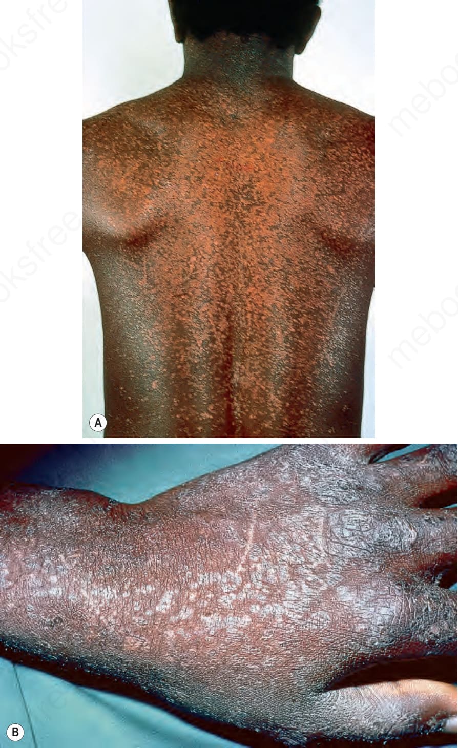
Fig. 18.27 Epidermodysplasia verruciformis: (A) innumerable small flat warts are present; (B) the dorsum of the hand is a commonly affected site. By courtesy of the Institute of Dermatology, London, UK.
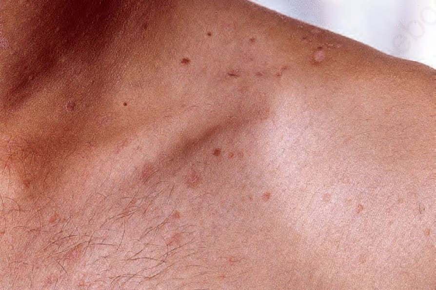
Fig. 18.30 Epidermodysplasia verruciformis: these scaly macules on the chest and axilla resemble pityriasis versicolor. From the collection of the late N.P. Smith, MD, the Institute of Dermatology, London, UK.