Plantar warts
Plantar warts
Clinical features Plantar warts occur on the sole of the foot; they are only slightly elevated and appear as a horny plug surrounded by a ring of hyperkeratotic skin
830 Infectious diseases of the skin
(Fig. 18.10). Often, they are covered with black dots representing thrombosed capillaries (Fig. 18.11).1 They are most common in children and are frequently seen over pressure points. Most plantar warts are caused by HPV1 and are painful; however, HPV4 may produce a confluent or mosaic pattern of similar small warts (‘mosaic plantar warts’) and these are painless (Fig. 18.12).2,3 They may also be seen on the palms and in the periungual region. There have been reports from Japan of unusual plantar warts produced by HPV60.4–6
The lesions may be nodular, ridged, or pigmented. A cystic variant has also been described.4–10 The cystic variant has the features of an epidermoid cyst and may rarely be multiple.11 Most are associated with HPV60, but an association with HPV57 has also been reported.12–14 Epidermoid cysts induced by HPV may also be seen outside acral locations.15,16 Pigmented
831 Plane warts
warts are caused by HPV4, 60, or 6517 and may contain fibrillar intracytoplasmic inclusion bodies.18 A case of a large plantar wart caused by HPV66 has been documented.19 A further subtype of HPV associated with palmoplantar warts is HPV63.8,20
Plantar warts usually regress within a few months in children, but may persist longer in adults. Rarely, chronic plantar warts may be associated with the development of verrucous carcinoma (carcinoma cuniculatum) (see Chapter 22).21,22
in the nuclei of these cells via electron microscopy (Fig. 18.16). Melanin granules are discernible within the cytoplasm of HPV60-induced pigmented plantar warts.
Regressive changes are the same as those described in common warts and consist of thrombosis of superficial blood vessels, necrosis, and a mixed inflammatory cell infiltrate.23 A recently described multiplexed PCR-based assay may have merit in both HPV genotyping and in monitoring treatment efficacy.24
Histologic features Plantar warts are almost entirely endophytic, with a central parakeratotic plug surrounded by multiple deep extensions of acanthotic epidermis (Fig. 18.13). The depth and complexity of these downgrowths have been likened to an anthill, giving rise to the term ‘myrmecia’. Vacuolation is more prominent in the plantar wart and, in the active growing phase, large eosinophilic (and to a lesser extent, basophilic) cytoplasmic inclusions are present, which represent disordered growth of giant keratohyalin granules (Fig. 18.14). The large eosinophilic cytoplasmic inclusions are usually seen in infections caused by HPV1 and to a lesser extent in those caused by HPV60 and HPV65.7 In warts induced by HPV4, the infected keratinocytes show prominent cytoplasmic vacuolar change with almost no keratohyalin granules. Intranuclear inclusions may also be evident (Fig. 18.15). HPV can be demonstrated
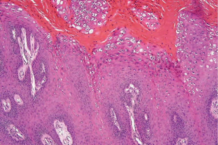
Fig. 18.7 Verruca vulgaris: large vacuolated cells with enlarged and irregular keratohyalin granules are characteristic.
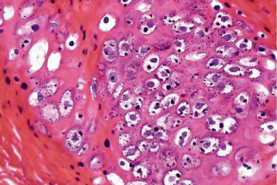
Fig. 18.8 Verruca vulgaris: high-power view of koilocytes.
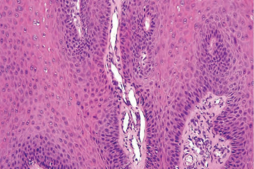
Fig. 18.9 Verruca vulgaris: the core of the papillary projection contains conspicuous dilated capillary loops.
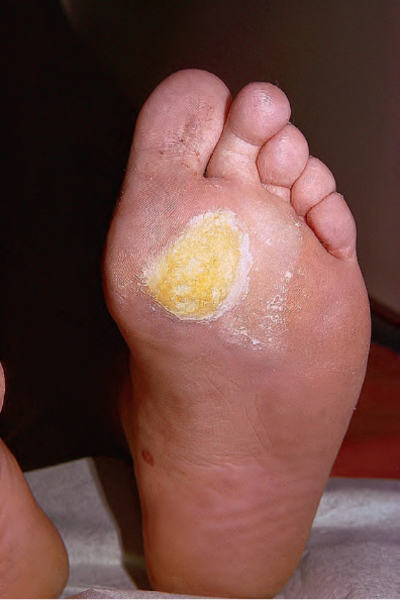
Fig. 18.10 Plantar wart: the lesion is flat and shows very marked hyperkeratosis. By courtesy of R.A. Marsden, MD, St George’s Hospital, London, UK.
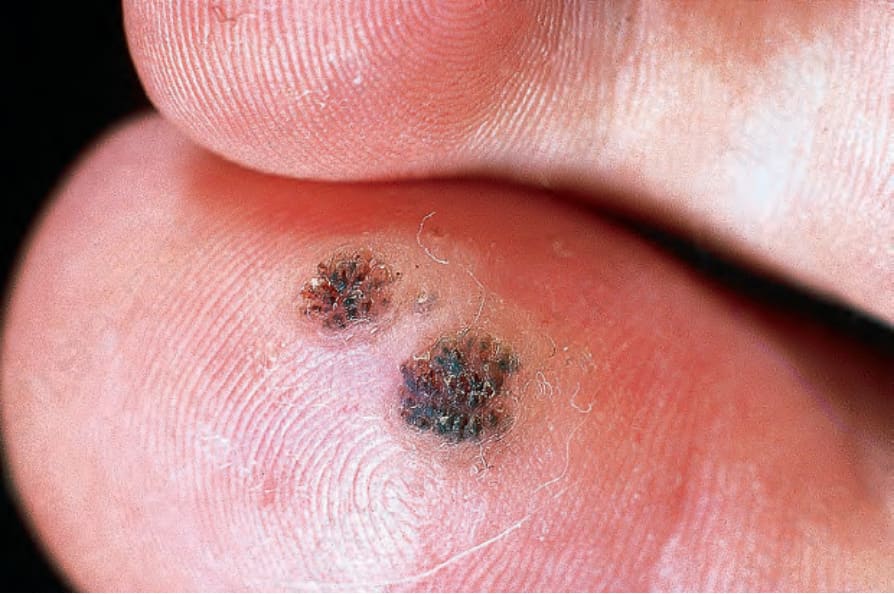
Fig. 18.11 Plantar wart: vascular thromboses as seen in these two lesions are common manifestations of involution. By courtesy of R.A. Marsden, MD, St George’s Hospital, London, UK.
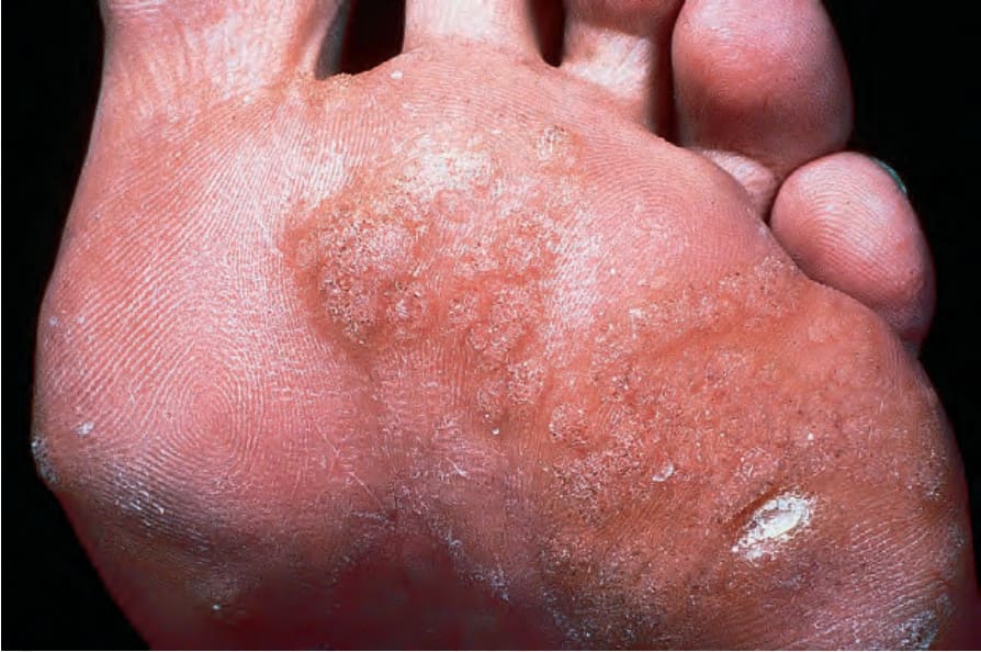
Fig. 18.12 Mosaic warts: here there are a large number of small warts. They are particularly resistant to therapy. By courtesy of the Institute of Dermatology, London, UK.
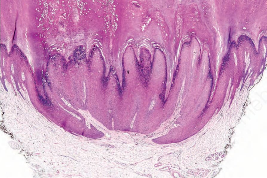
Fig. 18.13 Plantar wart: typical depressed, crateriform lesion containing a parakeratotic plug.
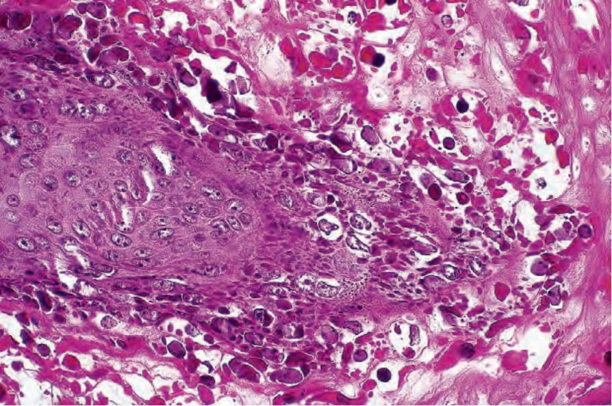
Fig. 18.14 Plantar wart: these eosinophilic keratohyalin granules are characteristic.
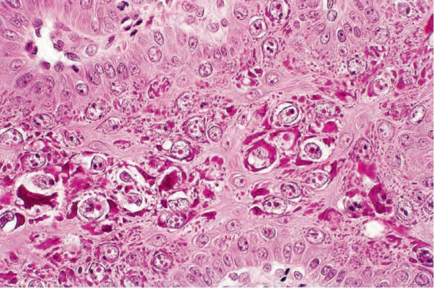
Fig. 18.15 Plantar wart: note the conspicuous intranuclear eosinophilic inclusions.
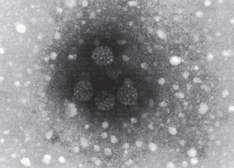
Fig. 18.16 Plantar wart: this honeycomb arrangement of HPV is characteristic. By courtesy of I. Chrystie, FIMLS, St Thomas’ Hospital, London, UK.