Hypergammaglobulinemic purpura
Hypergammaglobulinemic purpura
Clinical features Hypergammaglobulinemic purpura (of Waldenström) is a rare disorder that shows a marked female predilection and tends to affect the young and middle aged.1 Patients present with recurrent, symmetrical crops of purpura, particularly affecting the lower limbs although the arms and abdomen may also be involved (Fig. 16.132).2 Wearing tight-fitting garments, heat, and strenuous exercise may provoke lesions. The frequency of attacks is highly variable, ranging from several times a week to as infrequent as a single episode per year.3,4
The clinical findings are those of petechiae measuring from pinhead size up to several millimeters. Various symptoms may be experienced, including tingling, itching, burning, and pain. The petechiae resolve over the course of a few days to leave hyperpigmented macules. The purpuric attacks are
768 Vascular diseases
on chromosome 12 and involved in the biosynthesis of cholesterol and isoprenoid.1,2
The disease is most common in Europe and presents in childhood as recurrent episodes of high fever lasting for 3–7 days. Vaccinations and minor infections may be inciting events. Fever attacks are accompanied by abdominal symptoms, headaches, generalized lymphadenopathy, and arthralgias of large joints.3–6 Amyloidosis is a rare complication.3 Cutaneous manifestations include erythematous macules, papules, and nodules as well as urticarial lesions.7 Elevated IgD levels can be demonstrated in the majority of patients.3
Pathogenesis and histologic features The genetic defect results in inactivation of mevalonate kinase (MVK). The downstream effect is to increase production of IL-1 cytokines, resulting in a proinflammatory state.5,8,9 Skin biopsy findings are variable and most frequently show a mild acute vasculitis. Rarely, the features are reminiscent of Sweet syndrome, cellulitis, or erythema elevatum diutinum.4,7,10,11 Squamous syringometaplasia has also been reported.11
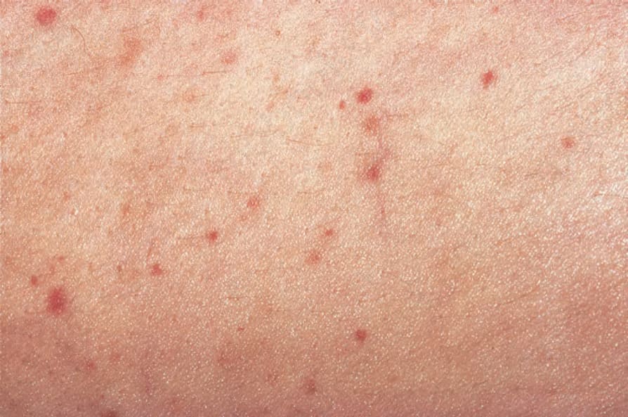
Fig. 16.132 Hypergammaglobulinemic purpura: scattered, tiny, purpuric lesions. By courtesy of J. Newton-Bishop, MD, St Thomas’ Hospital, London, UK.