Paraneoplastic vasculitis
Paraneoplastic vasculitis
Clinical features Occasionally, cutaneous vasculitis is a marker of an underlying systemic malignancy (Table 16.13).1–12 Most commonly the vasculitis is associated with hematological malignancies.12 There are also reports of an association with solid tumors.1,4,6,12–17 Vasculitis has nevertheless been reported in patients with carcinoma of the kidney, breast, ovary, lung, nasopharynx, stomach, small bowel, colon, prostate, and thyroid, and chondrosarcoma. Some of these associations may represent coincidence. Leukocytoclastic vasculitis is the most common pattern of vasculitis associated with malignancy but large-vessel vasculitis may also be seen. Of interest, vasculitis can be present at the time of initial diagnosis and also herald the onset of relapse.4 Solid tumors and hematological malignancy have been associated with IgA vasculitis.7,10–12 One study found that nearly a third of adults with Henoch-Schönlein purpura had an associated malignancy.10 For this reason, the authors concluded that physicians should suspect underlying malignancy in older patients (especially men of more than 40 years) with IgA vasculitis.10
In contrast to solid tumors, there does appear to be a more definitive relationship between cutaneous vasculitis and hematological and lymphoreticular neoplasms including hairy cell leukemia, acute and chronic myeloid leukemia, multiple myeloma, and non-Hodgkin lymphoma.1
A
In general, patients present with the features of leukocytoclastic vasculitis, and occasionally arthralgia or arthritis is evident. Cutaneous manifestations include maculopapular eruptions, purpura, urticaria, peripheral ulcers, and gangrene.12,18 Although vasculitis is seen in patients with a spectrum of hematological malignancies, hairy cell leukemia is particularly associated with leukocytoclastic vasculitis and a polyarteritis nodosa-like picture, including systemic lesions.4–6 In one study of 42 patients with hairy cell leukemia and vasculitis, 21 also had leukocytoclastic vasculitis and 17 had polyarteritis nodosa.4 In addition, four patients had direct infiltration of vessel walls by leukemic cells. Hodgkin lymphoma has occasionally been linked to
B
• Migratory superficial thrombophlebitis
• Deep venous thrombosis
• Nonbacterial thrombotic endocarditis
• Anticardiolipin antibody syndrome
• Embolic features associated with atrial myxoma
• Raynaud phenomenon
• Erythema nodosum
• Hyperviscosity syndrome
• Cryoglobulinemia
• Lambda light chain vasculopathy
• Cutaneous vasculitis
• Systemic vasculitis
Reproduced with permission from Mertz, L.E. and Conn, D.L. (1992) Current Opinion in Rheumatology, 9, 39–46.
754 Vascular diseases
erythema nodosum, and myelodysplasia has been found in conjunction with leukocytoclastic vasculitis.19,20 Multiple myeloma is particularly associated with nonthrombocytopenic purpura.21 It has been documented that lymphocytic vasculitis is a relatively common form of paraneoplastic vasculitis associated with lymphoproliferative disorders.22
It is important to note that these vasculitic phenomena may antedate the clinical manifestations of the underlying malignancy. In one retrospective study from a single tertiary care institution, the diagnosis of vasculitis in all 16 cases preceded the diagnosis of malignancy.12 Therefore, patients with an unexplained vasculitic rash should be investigated with this in mind.2,12 In hairy cell leukemia, vasculitis often follows splenectomy.5,18
Differential diagnosis The precise terminology that is preferred by the dermatopathologist is probably not important. More significant than the nosological nuances is rendering a report that alerts the clinician to the possibility that the patient may have underlying systemic disease, and when such lesions are encountered appropriate clinical evaluation is necessary.
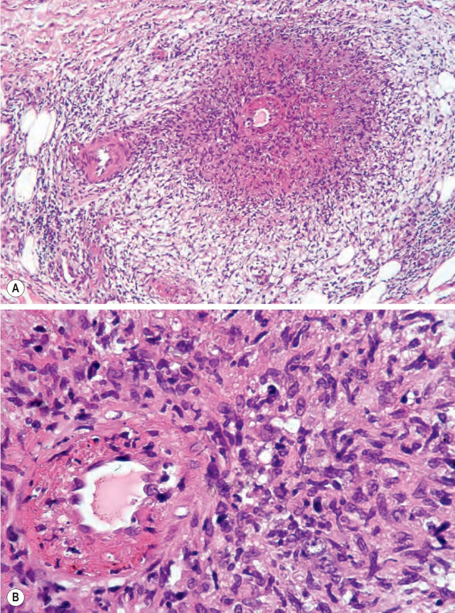
Fig. 16.99 (A, B) Tuberculous vasculitis: this patient with miliary tuberculosis presented with ischemic cutaneous lesions. Note the granulomatous inflammation.
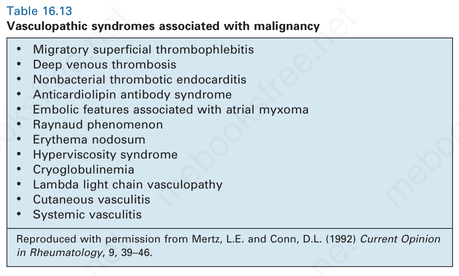
Table 16.13 Vasculopathic syndromes associated with malignancy