Infantile acute hemorrhagic edema
Infantile acute hemorrhagic edema
Clinical features Infantile hemorrhagic edema is a form of leukocytoclastic vasculitis that is mostly seen in newborns but has also been described in the first 3 years of life and occasionally in older children.1–11 The disease is usually limited to the skin but mucosal involvement may be an additional feature.10 Transient renal involvement with microscopic hematuria and proteinemia, hypocomplementemia, abdominal pain, and elevated transaminases are exceptional additional findings.12,13 It frequently follows vaccination or infection including otitis, upper respiratory tract infection, or conjunctivitis.1,10,11 An association with cytomegalovirus, herpes simplex virus-1, or rotavirus infection has been documented.14–16 Since many children had received antibiotics for infection prior to development of lesions, a subset of cases may represent reaction to medication.1 This disease has a peak incidence in the winter months.
Skin lesions are widely distributed, and often involve the head and neck, and limbs. They present as purpuric lesions that often have a rosette or targetoid configuration.2,3 The cheeks and ears seem to be sites of predilection.3,11 Resolution within a few weeks is typical and recurrences are not reported.1,11,17
An elevated ESR and leukocytosis are usually present.
Pathogenesis and histologic features The pathogenesis of infantile hemorrhagic edema is unknown; however, it is likely that the disease is immune mediated. Biopsy shows features of leukocytoclastic vasculitis with variable fibrinoid necrosis.3
Differential diagnosis Some authors consider infantile hemorrhagic edema to be a variant of IgA vasculitis. Others do not agree, arguing that the absence of perivascular IgA on immunofluorescence staining, absence of systemic involvement in most patients, and the benign clinical course do not support this view.3,18 However, a very interesting link between the two diseases has been postulated.19 Goraya and Kaur note that, since the IgA immune system in infants is immature, if acute hemorrhagic edema were related to IgA vasculitis, the patient would be incapable of mounting an IgA-mediated immune response and this would explain the lack of IgA on immunofluorescence studies in the majority of patients.19 Clearly, further study of this disorder is necessary to elucidate its pathogenesis and to clarify its nosological position in the classification of leukocytoclastic vasculitis.
often last 24–72 hours.9 Patients commonly complain of pruritus, burning, or pain. The frequency of cutaneous symptoms varies considerably, from daily to monthly.
Joint pain, stiffness, and swelling, particularly of the hands, elbows, feet, ankles, and knees, are seen; however, frank arthritis is extremely rare.10 Hypocomplementemia, which correlates with systemic involvement, is a feature in many patients.4,7,8,11–13 Patients with hypocomplementemia may have musculoskeletal involvement, ocular involvement, pulmonary involvement, and gastrointestinal involvement in decreasing order of frequency.13 Proteinuria and hematuria may also be noted. Rarely, patients develop focal or diffuse proliferative glomerulonephritis. Crescentic glomerulonephritis, mesangial glomerulonephritis, and membranous nephropathy have also been documented.8,14–19 Gastrointestinal symptoms can include abdominal pain, nausea, vomiting, and diarrhea, and an associated peripheral neuropathy has been reported.13,20
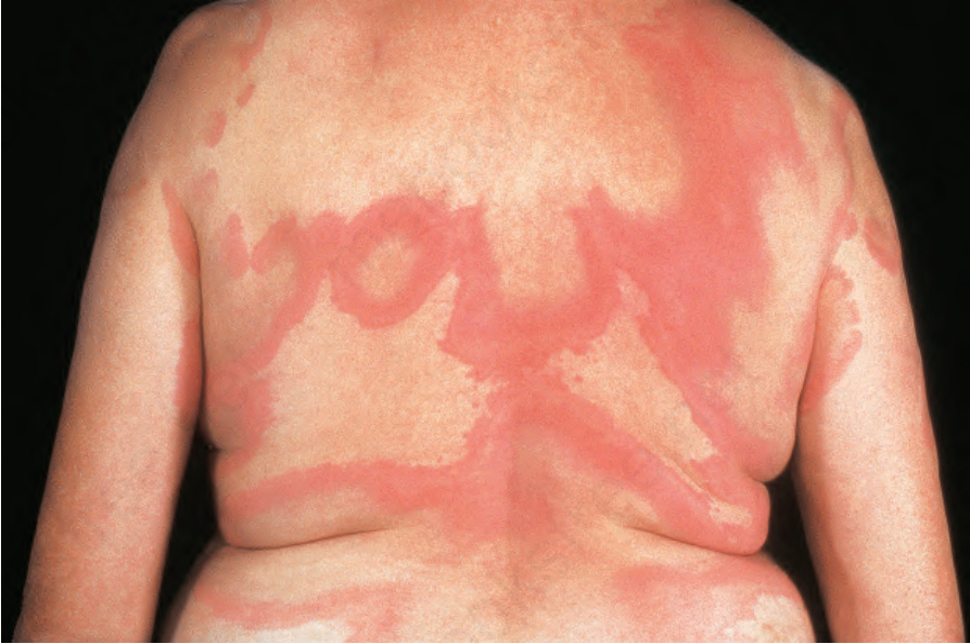
Fig. 16.23 Urticarial vasculitis: this very large lesion has developed a bizarre outline due to central clearing. By courtesy of the Institute of Dermatology, London, UK.
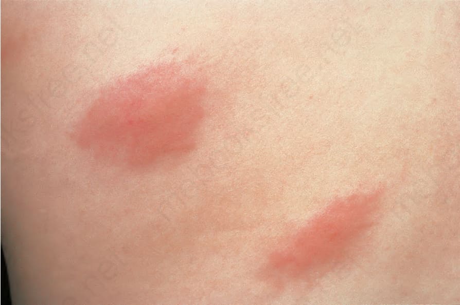
Fig. 16.24 Urticarial vasculitis: close-up view. By courtesy of the Institute of Dermatology, London, UK.