Esthetic microimplants
Esthetic microimplants
Cosmetic dermatology is a rapidly evolving industry. The demand for ‘nonsurgical’ rejuvenation has seen a rise in the number of implants and injectable materials available to patients. Unfortunately, inflammatory reactions to these agents occur.
There are several categories of fillers which may be permanent or resorbable (Table 14.2). Fillers are either polymers, which function as volumizers, creating effect by taking up space, or a combination of degradable polymer and microparticles.1 The microparticles serve as a lattice upon which the host’s response (often collagen induction) contributes partially or completely to the filler’s effect. The microparticles may or may not be degraded over time.
As a consequence of the ban on silicone, bovine collagen was developed. There is a 1–3% rate of allergic reactions, despite double skin testing.4 Many such reactions are localized erythema and edema which occur in the first weeks to months after injection, although recurrent inflammation, years later, has been described. Other potential complications include localized glabellar necrosis, granulomata, and abscess formation.5 Granulomata present as erythematous nodules at the site of treatment and are seen within a few months following injection. Sometimes, lesions are seen years later. Abscesses typically occur weeks to months after injection.4,5 Polymethyl-methacrylate microspheres and bovine collagen (Artecoll, Arteplast, and Artefill) is a temporary filler that uses a solution of bovine collagen as a carrier. Patients may rarely develop telangiectasias, hypertrophic scarring, allergic reactions, and granuloma formation.5
Chemical ingredient Product name(s)
Resorbable Bovine collagen Zyderm, Zyplast
Human collagen Cosmoderm, Cosmoplast, Cymetra
Porcine collagen Evolence
Hyaluronic acid (nonanimal) Restylane, Captique, Perlane, Juvederm
677 Esthetic microimplants
nongranulomatous. The etiology of the granulomatous response is unclear. There is speculation that it could be secondary to impurities related to the fermentation process in the nonanimal-derived form.13 Additional side effects to hyaluronic acid include sterile abscesses, hyperpigmentation, and a livedoid, reticular pattern in the area of injection.8
Hydroxyethylmethacrylate/ethylmethacrylate fragments and hyaluronic acid (DermaLive and DermaDeep) is a permanent filler. It can result in edema, induration, and granulomatous inflammation.5
Foreign-body granulomata have also been seen following injection with poly-L-lactic acid (Newfill, Sculptra) and polyacrylamide gel (PAAG; Aquamid).4–6 Hydroxy-polyethylene/hydroxyl-polypropylene (Profill) is also associated with lipoatrophy.4
Hyaluronic acid (animal) Hylaform, Hylaform plus
Poly-L-lactic acid Newfill, Sculptra
Hydroxy-polyethylene/ hydroxyl-polypropylene
Profill
Calcium hydroxylapatite Radiesse, Radiance
Permanent Bovine collagen/polymethyl methacrylate microspheres (PMAA)
Artecoll, Arteplast, Artefill, Metacril
Hyaluronic acid/ polyhydroxyethylmethacrylate (HEMA)/ethylmethacrylate (EEMA)
DermaLive, DermaDeep
Calcium hydroxylapatite is a resorbable filler of calcium hydroxylapatite microspheres that stimulate the endogenous production of collagen. Occasional granulomatous reactions occur.5
Polytetrafluoroethylene (Gore-Tex) has been used for lip augmentation and has resulted in ulcerated nodules with pustules.14
Immediate-type reactions such as erythema, edema, and bruising have also been described with other fillers, including hydroxy-polyethylene/ hydroxyl-polypropylene (Profill), polyacrylamide-containing products, and poly-L-lactic acid.4,14
Infection is a potential complication following any cosmetic procedure.14 Early infections, occurring within 2 weeks, are typically secondary to bacteria such as S. aureus or Streptococcus.3,14 Later infections may be due to atypical mycobacteria such as Mycobacterium chelonae, Mycobacterium fortuitum, and abscesses.14
Silicone (dimethyl polysiloxane) Silikon 1000, Silskin, Adatosil 5000
Polydimethylsiloxane/polyvinyl pyrrolidone
Bioplastique
Polyacrylamide gel (PAAG) Aquamid
Polyacrylamide and polyvinyl acid Evolution
Polyalkylimide gel Bio-Alcamid
Polytetrafluoroethylene Gore-Tex
Histologic features Silicone nodules contain multiple vacuoles in the dermis, subcutis, and skeletal muscle.3,4,13 Vacuoles are variable in size and shape and resemble Swiss cheese. These are actually empty spaces where silicone was lost during processing. They are surrounded by giant cells and histiocytes which contain intracytoplasmic vacuoles.15 The latter may mimic lipoblasts. Additionally, impurities in the silicone result in birefringent foreign material within giant cells.3–5 Surrounding fibrosis may also be noted.3 The silicone particles in polydimethylsiloxane/polyvinyl pyrrolidone are too large to be phagocytosed by histiocytes.1 The material induces a foreign body giant cell reaction and fibrosis. The particles are identified as translucent, jagged structures within cystic spaces.4,5
Bovine collagen is distinguished from human collagen by its lighter eosinophilic color, thicker bundles, and more amorphous, acellular appearance.4,5 When recurrent inflammatory reactions develop in hypersensitive patients, there is a perivascular mononuclear cell infiltrate with a mixture of neutrophils, mononuclear cells, and eosinophils within implanted collagen.4,15
Early granulomatous nodules are composed of a mixture of mononuclear cells, giant cells, eosinophils, neutrophils, and plasma cells surrounding, but not infiltrating, the collagen implant.4 Later lesions have a denser and deeper infiltrate. Birefringent material is not seen. In contrast, nodules due to polymethyl-methacrylate microspheres and bovine collagen (PMAA) (Artecoll) contain a diffuse and nodular granulomatous infiltrate which surrounds cystic spaces, mimicking fat cells and with a Swiss cheese appearance.3,5,15 Located in the center of the spaces are nonbirefringent, round, translucent, well-circumscribed foreign bodies (Fig. 14.123). This foreign material is the PMMA microsphere, which also induces surrounding fibrosis. The fibrosis contributes to the filling effect. Hyaluronic acid with polyhydroxyethylmethacrylate (HEMA)/ethylmethacrylate (EEMA) (DermaLive, DermaDeep) reactions are very similar histologically to Artecoll, as they also contain methacrylate microspheres, but the foreign material has a more polygonal appearance.3,5,15 Calcification of the foreign material has been described with DermaLive.1 Asteroid bodies may be present in both reactions.15
Human-derived bioengineered collagen was developed as an alternative to bovine collagen. Most reactions consist of localized bruising at sites of injection, but rare granulomatous reactions have been reported.5
Hyaluronic acid is a temporary filler commonly used to correct facial creases and wrinkles. There are two types of hyaluronic acid fillers: nonanimal, derived from fermentation of bacteria (Streptococcus equi), and animal, derived from chicken combs.5–7 Hypersensitivity reactions, although rare, have been reported in up to 0.8% of patients.4,8 Injection site reactions are most common, with temporary erythema, edema, and bruising within the first 14 days following injection.9 Delayed reactions are less common and present as erythematous, firm nodules along the sites of injection weeks to months later.6–12 There are two types of nodules: granulomatous and
Abscesses seen as a consequence of collagen implants show a dense neutrophilic infiltrate with admixed plasma cells, histiocytes, and mononuclear cells.4 Giant cells surround implanted collagen, and granulomata may be present.
Nongranulomatous inflammatory nodules related to hyaluronic acid injection are characterized by a superficial and deep perivascular and
678 Cutaneous adverse reactions to drugs
A
B
periadnexal infiltrate of mononuclear cells with several eosinophils.13 Implanted hyaluronic acid is not seen. In contrast, the granulomatous nodules contain a striking nodular infiltrate of foreign body giant cells, histiocytes, and eosinophils surrounding pools of basophilic foreign material which stain with Alcian blue (pH 2.7).4,14 Polyacrylamide gel (Aquamid) has similar features, but is distinguished from injected hyaluronic acid by the presence of necrotic tissue admixed with the basophilic foreign material.5,14
Fig. 14. 125 Reaction to Bio-Alcamid: the central necrobiosis with a surrounding rim of histiocytes shows a striking resemblance to granuloma annulare.
Poly-L-lactic acid (Newfill, Sculptra) nodules are granulomatous and are distinguished by spiky, long translucent bodies within giant cells (Fig. 14.124).4,14 They are irregular in shape and birefringent.
Reactions to Bio-Alcamid (polyalkylimide gel) display palisading granulomas with scattered giant cells and a central area of amorphous material that may mimic deep granuloma annulare or rheumatoid nodule (Fig. 14.125).5,15
Ulcers caused by polytetrafluoroethylene (Gore-Tex) demonstrate variably sized threads of material surrounded by neutrophils and granulation tissue (Fig. 14.126).14
679 Aluminum granuloma
Infectious lesions demonstrate variable findings depending on the causative organism. Mycobacterial infections may present as granulomatous nodules, and appropriate tissue stains and culture are necessary.
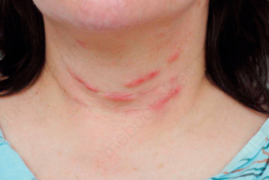
Fig. 14.122 Restylane nodules: linear arrangement of nodules along the neck creases.
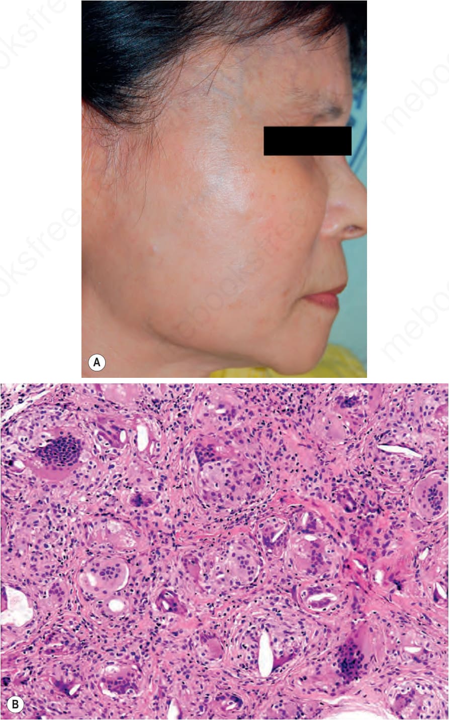
Fig. 14.123 (A) Clinical reaction to poly-L-lactic acid filler. (B) Reaction to a poly-L-lactic acid with granulomas containing spiky translucent material. By courtesy of Chao-Kai Hsu, MD, Sheau-Chiou Chao, and Julia Yu-Yun, National Cheng Kung University Hospital, Tainan, Taiwan.
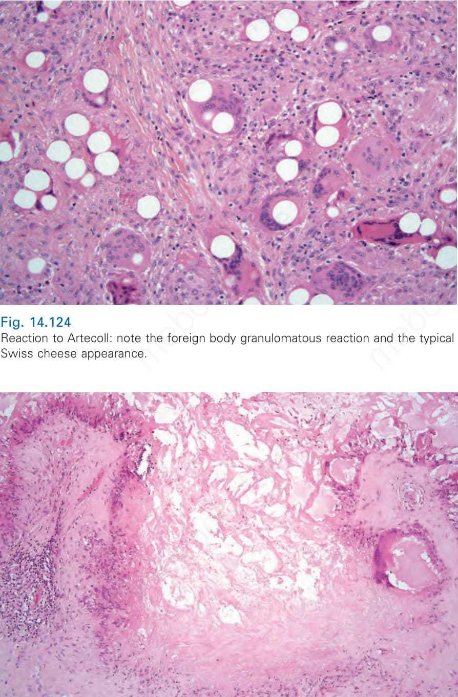
Fig. 14.124 Reaction to Artecoll: note the foreign body granulomatous reaction and the typical Swiss cheese appearance.
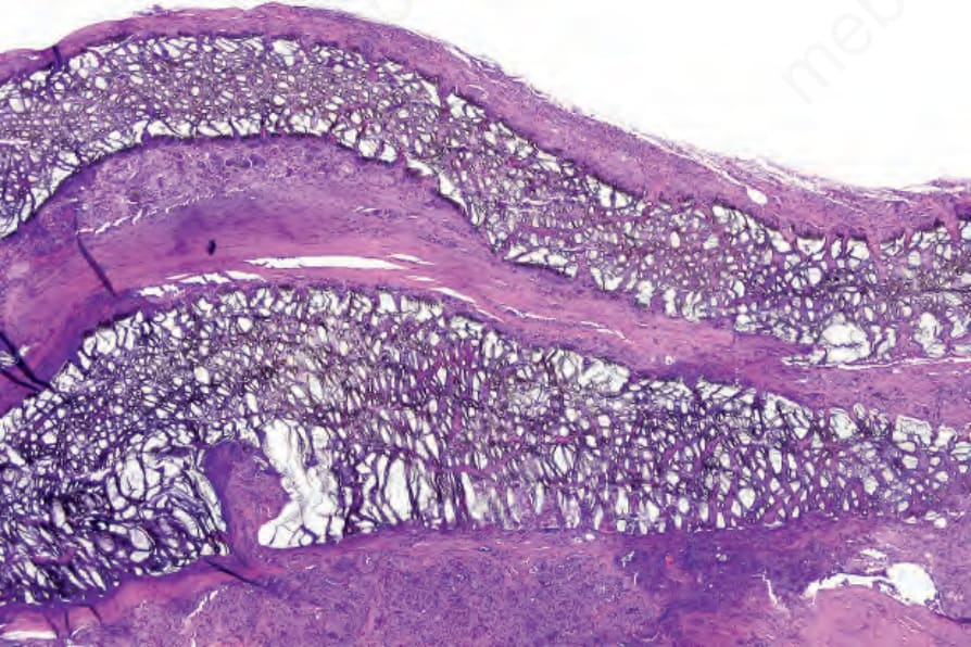
Fig. 14.126 Reaction to Gore-Tex: the mesh is surrounded by dense fibrous tissue.
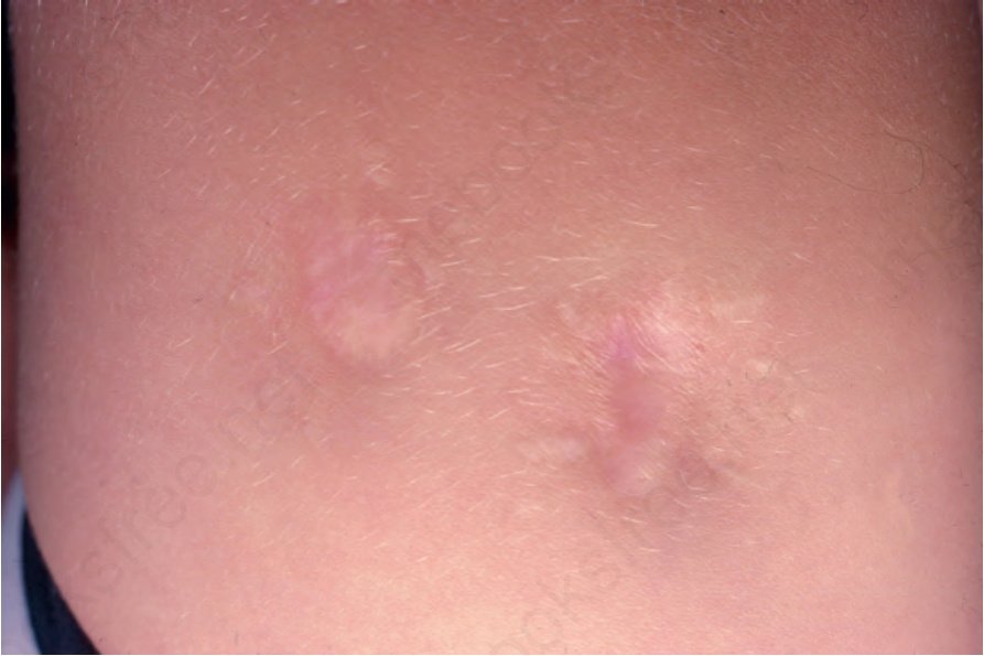
Fig. 14.127 Aluminum granuloma: multiple depressed nodules with scarring are evident. From the collection of the late N.P. Smith, MD, Institute of Dermatology, London, UK.
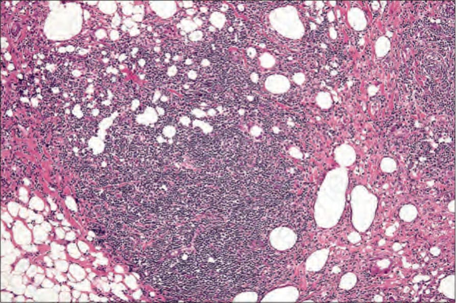
Fig. 14.128 Aluminum granuloma: there is a dense inflammatory cell infiltrate within the subcutaneous fat.
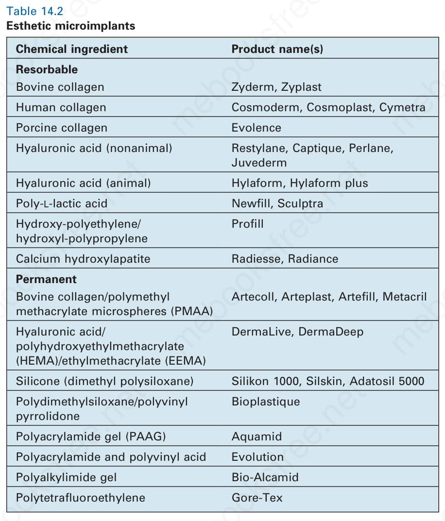
Table 14.2 Esthetic microimplants
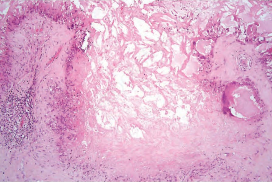
Fig. 14-125 (caption embedded in image / 圖說烘焙於圖內)