Generalized myxedema
Generalized myxedema
B
Clinical features Generalized myxedema occurs as a consequence of severe hypothyroidism. Patients with myxedema may appear pale yellow due to the combined effects of edema, anemia, and carotenemia.1,2 The last, which is due to defective conversion of beta-carotene to vitamin A in the liver, is seen particularly on the palms, soles, and in the nasolabial folds.3 Rarely, the color change is generalized.4 The skin is cool, dry, coarse, waxy, and puffy, especially around the eyes and cheeks, and the hands and feet may show nonpitting edema (Fig. 13.162).3,5–7 The face is often expressionless. Eccrine and sebaceous gland secretions are reduced and this may result in xerosis, an ichthyotic appearance, or asteatotic eczema.4 Hyperkeratosis over bony prominences resembling avitaminosis A is also sometimes evident.8 Alopecia is a common finding and the outer third of the eyebrows is typically affected. There is usually thinning of the beard and sexual hair in addition to loss of the scalp hair. Myxedema is associated with a greatly increased percentage of hair follicles in the telogen phase.9 The rate of hair growth is
614 Degenerative and metabolic diseases
also diminished. Residual hair is dry, coarse, and brittle.3 The nails often become thin, brittle, and striated.1 Additional cutaneous manifestations have included pruritic papular lesions, purpura and ecchymoses, impaired healing, generalized follicular mucinosis, and multiple focal cutaneous mucinoses.1,5,9–11 Oropharyngeal and laryngeal involvement is common and many patients are hoarse. Patients with myxedema have an increased risk of developing hyperlipidemia with resultant eruptive and tuberous xanthomata (Fig. 13.163).
A
Histologic features The epidermis may show mild hyperkeratosis with occasional follicular plugging.3 Most frequently, the dermal changes are subtle and nondiagnostic. However, in cases of greater severity, there is slight swelling and separation of the collagen bundles with edema, and special stains show that small quantities of mucin are present within the dermis and occasionally in the subcutaneous fat.12 Fibroblastic proliferation is not a feature of generalized myxedema.13

Fig. 13.160 Mucinosis: (A) actively secreting fibroblasts contain abundant rough endoplasmic reticulum; (B) numerous intracytoplasmic vesicles containing amorphous material are commonly present.
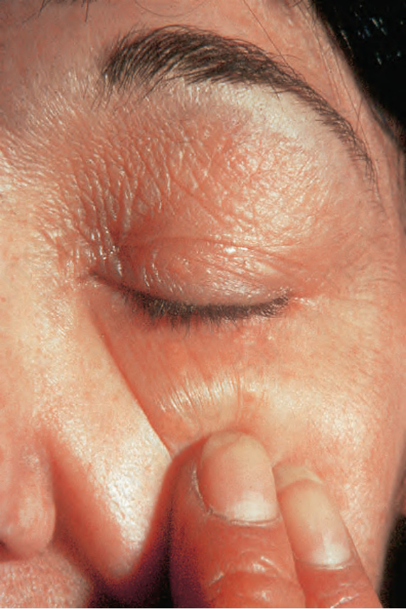
Fig. 13.162 Generalized myxedema: note the waxy infiltrated plaques on the eyelid. By courtesy of R.A. Marsden, MD, St George’s Hospital, London, UK.
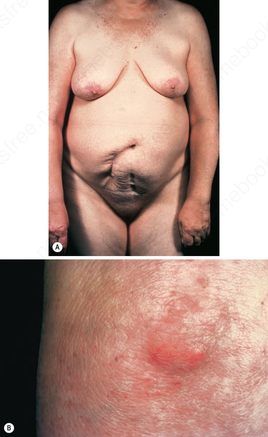
Fig. 13.163 (A, B) Generalized myxedema: this patient has widespread xanthomata. By courtesy of the Institute of Dermatology, London, UK.