The mucinoses
The mucinoses
The mucinoses are a group of conditions in which accumulation of acid glycosaminoglycans (mucin), particularly hyaluronic acid and to a lesser extent chondroitin (-4 and -6) sulfate and heparin, occurs either diffusely or focally in the dermis (Table 13.6).1–6 Mucinosis also may occur as a secondary phenomenon in dermatoses such as lupus erythematosus, scleroderma, dermatomyositis, Degos disease, granuloma annulare, and chronic graft-versus-host disease.5,7,8 In this chapter, however, only primary cutaneous mucinoses are the focus.
The glycosaminoglycans, which are secreted by fibroblasts, are constituents of normal cell membranes and connective tissue. This substance is usually secreted in only small amounts by fibroblasts. It is not clear why mucin production is increased in pathological states. Although the cause is probably multifactorial, it has been suggested that cytokines and/or immunoglobulins and unidentified factors in the serum of affected patients can induce synthesis of glycosaminoglycans.5,9–11 Cytokines that play an important role in this process include tumor necrosis factor, interleukin-1, and transforming growth factor beta (TGF-β).5,12,13 Actively secreting fibroblasts have a characteristic stellate shape and contain intracytoplasmic secretory vesicles; their presence in sections should therefore prompt a careful search for mucin deposition (Figs 13.158–13.160).
Diffuse
lichen myxedematosus – generalized form (scleromyxedema) scleredema reticular erythematous mucinosis generalized myxedema pretibial myxedema hereditary progressive mucinous histiocytosis Focal
lichen myxedematosus – discrete papular form acral persistent papular mucinosis papular and nodular mucinosis associated with lupus erythematosus self-healing juvenile cutaneous mucinosis cutaneous mucinosis of infancy cutaneous focal mucinosis myxoid cyst Follicular
follicular mucinosis
613 The mucinoses
Hyaluronic acid stains with colloidal iron (blue–green), Alcian blue at pH 2.5 (blue) (but not at pH 0.4), and mucicarmine (red) but it is negative for PAS. It also stains metachromatically with toluidine blue, methylene blue, and thionine.14 Sulfated acid mucins stain with Alcian blue at pH 0.5 and aldehyde-fuschin.2 Hyaluronic acid absorbs enormous amounts of water, which accounts for the induration and thickening common to this group of conditions.15
Routine fixation and processing results in an anhydrous state so that mucin presents as basophilic strands and granules in hematoxylin and eosin stained sections.3 In normal skin it is found particularly around appendages and the vasculature (Fig. 13.161). In the cutaneous mucinoses the deposits are hyaluronidase sensitive because most of the mucin present is hyaluronic acid. The excessive mucin disrupts the collagen fibers, giving them a frayed appearance. In general, with the exception of scleromyxedema, there is considerable histologic overlap within this group of conditions. Diagnosis depends considerably upon clinical features and the results of biochemical investigations.15
A
There are five major mucinoses:
• generalized myxedema,
• pretibial myxedema,
• lichen myxedematosus,
• reticular erythematous mucinosis,
• scleredema. Follicular mucinosis is considered in Chapter 29.
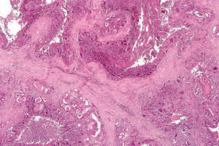
Fig. 13.155 Tumoral calcinosis: this low-power view shows a dense hyalinized stroma with numerous cystic cavities containing necrotic and calcified debris.
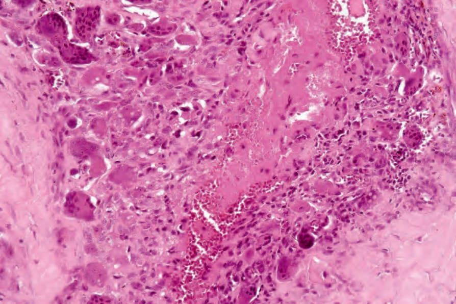
Fig. 13.156 Tumoral calcinosis: early lesions characteristically show a histiocytic and giant cell palisade around eosinophilic, degenerate connective tissue.
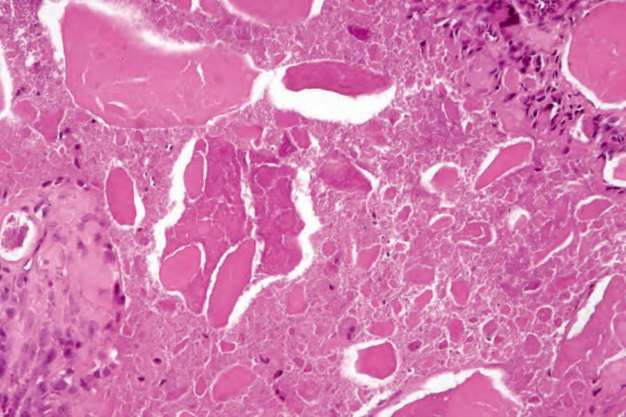
Fig. 13.157 Tumoral calcinosis: in older lesions, calcified deposits lie within lacunae.
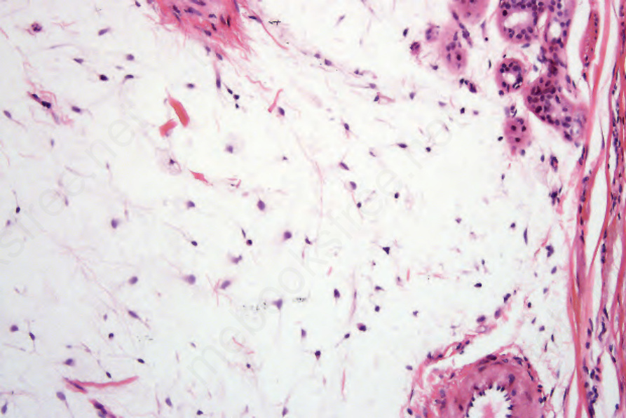
Fig. 13.158 Myxoma: Note abundant stromal mucin.
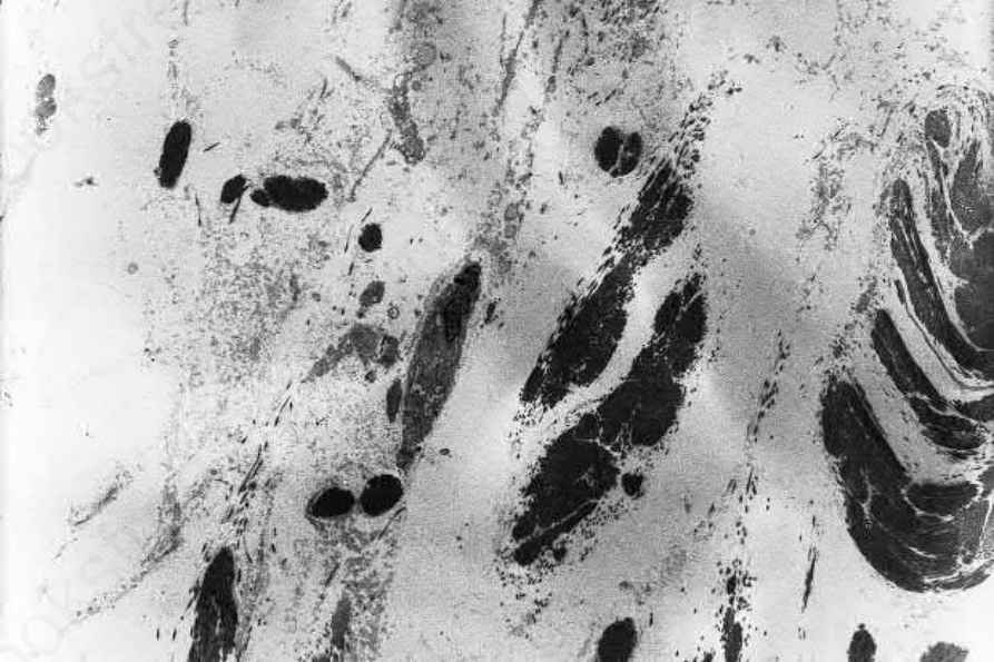
Fig. 13.159 Mucinosis: this electron micrograph from a patient with acral persistent papular mucinosis shows collagen bundles widely separated by a faintly electron-dense granular deposit.
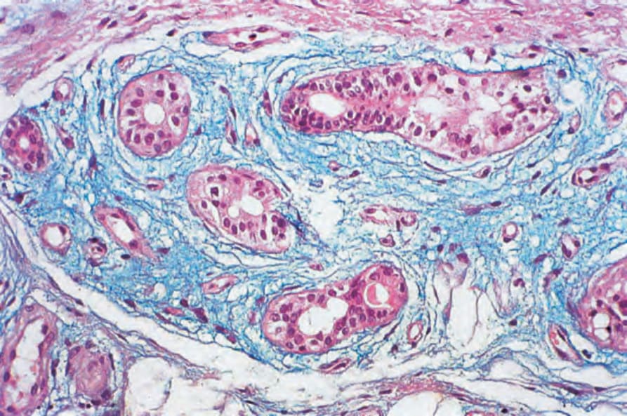
Fig. 13.161 Eccrine sweat gland: this section of normal skin from the sole of the foot shows abundant dermal mucin when stained with Ehrlich hematoxylin.
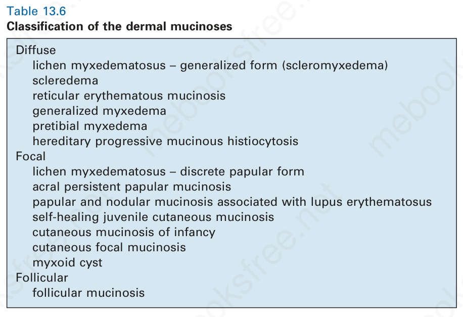
Table 13.6 Classification of the dermal mucinoses