Pellagra
Pellagra
Clinical features Pellagra develops as a consequence of deficiency of nicotinic acid (niacin, vitamin B3) or its precursor tryptophan.1–3 The cause may be dietary. It has traditionally been associated with high consumption of corn.3 In developed countries it is most frequently observed in alcoholics, in those living in conditions of socioeconomic deprivation, and in patients with anorexia nervosa or malabsorption due to extensive gastrointestinal disease (e.g., partial gastrectomy, gastroenterostomy, and Crohn disease).1,4 A severe case of cytomegalovirus colitis in an immunocompetent patient has also been associated with pellagra.5 It is sometimes also a feature of the carcinoid syndrome because the tumor cells consume available tryptophan to produce serotonin.5,6 Pellagra occasionally develops after therapy with a number of drugs, including isoniazid, 6-mercaptopurine, 5-fluorouracil, ethionamide, and phenobarbital.6–13 It can occur in Hartnup disease and in association with defects in the metabolism of tryptophan.14 Rare cases have also been associated with megaduodenum, congenital duodenal diaphragm, and Sjögren syndrome.15–17 Pellagra is particularly prominent in parts of Africa and Asia where nutritional deficiencies are prevalent.1 A rare case has been described in association with the intake of alternative medicines.18 A further report describes an association with amyloidosis secondary to multiple myeloma.19 A pellagra-like eruption partially responsive to niacin associated with xeroderma pigmentosum/Cockayne syndrome complex has been reported.20
features sometimes present include cheilosis, glossitis, angular stomatitis, and oral or perianal sores.3,7
Gastrointestinal disease in pellagra manifests as nausea, vomiting, abdominal pain, gastritis, and diarrhea. Neurological involvement evolves with headache, depression, and ataxia initially then more severe symptoms of disorientation, delirium, coma and eventually death.3
The classic triad of pellagra is ‘dermatitis, diarrhea, and dementia’. The skin eruption of pellagra is photosensitive in nature. An initial painful sunburn-like erythema subsides to leave a dusky brownish discoloration with a dry scaly appearance (Figs 13.139 and 13.140). Blisters may sometimes be evident. The eruption is typically sharply demarcated, symmetrical, and occurs on the backs of the hands (most commonly), the forearms, the knees, central chest, neck, and face.7 The thickened skin around the photo-exposed skin of the neck typically resembles a necklace (Casal necklace). Other
Histologic features The appearances in pellagra are usually non-specific. There is hyperkeratosis, parakeratosis, and acanthosis associated with increased melanin pigmentation and, in early lesions, keratinocyte vacuolation in the upper reaches of the epidermis.7,8 Telangiectasia and a perivascular chronic inflammatory cell infiltrate in the upper dermis may also be evident. Older lesions sometimes show epidermal psoriasiform hyperplasia.21 In some instances the histology can resemble that of necrolytic migratory erythema and acrodermatitis enteropathica.
Differential diagnosis The diagnosis is very much dependent upon clinicopathological correlation, particularly in those cases that resemble necrolytic migratory erythema and acrodermatitis enteropathica.
607 Calcinosis cutis
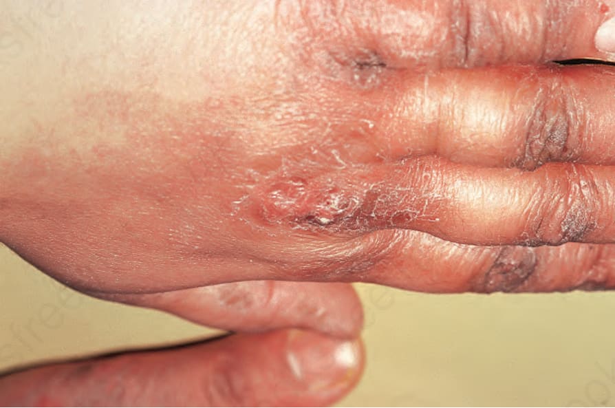
Fig. 13.139 Pellagra: scaling and hyperpigmentation are present on the dorsal aspect of the knuckles and fingers. By courtesy of the Institute of Dermatology, London, UK.
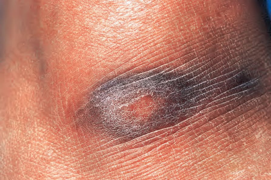
Fig. 13.140 Pellagra: close-up view of hyperpigmentation and scaling. By courtesy of the Institute of Dermatology, London, UK.
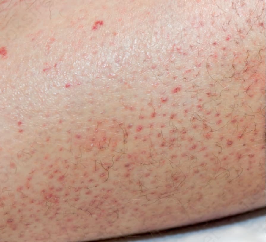
Fig. 13.141 Scurvy: the hairs have a coiled, corkscrew shape and there is perifollicular hemorrhage. By courtesy of Melissa Piliang, MD, Cleveland Clinic, Cleveland, USA.