Exogenous ochronosis
Exogenous ochronosis
Clinical features Deposition in the skin of an identical pigment to that seen in alkaptonuria may occur as a result of the application of phenol (carbolic acid) to leg ulcers, therapy with resorcinol and picric acid, the oral and intramuscular administration of antimalarials such as chloroquine, and the application to dark skin of bleaching creams containing hydroquinone, most often in black women.29–42 Antimalarials result in slate-gray pigmentation affecting the knees, face, palate, and subungual regions.43 In hydroquinone-induced ochronosis, lesions occur particularly over bony prominences such as the forehead, temples, nose, and lower jaws and also on the sides of the neck (Fig. 13.136).33 Time to onset of lesions is approximately 6 months. The first stage is characterized by erythema and mild hyperpigmentation.30 Subsequently, the hyperpigmentation intensifies and patients develop widespread ‘caviar-like’ black papules; cutaneous atrophy and colloid milia may also occur.44 In longstanding disease, nodules develop.30,45–47 Hydroquinone-induced ochronosis is a major problem in the black population of South Africa. In one series the prevalence among users of skin lighteners was almost 70%.34 The reason for the high incidence of ochronosis in this population is not entirely clear but is thought to be due in part to high concentrations of hydroquinone used in their products and the synergistic
Osteoarticular involvement – which is particularly evident in the knees, shoulders, and hips, and in advanced cases the vertebral column – is characterized by pigmentation of the articular cartilage, synovium, and capsule associated with fibrillation, fragmentation, calcification, and erosion.13,14 Osteoarthritis may also be evident and chronic non-specific synovitis is commonly present. Cardiovascular involvement occurs in up to 50% of
605 Ochronosis
effect of multiple compounds used in combination with hydroquinone such as mercury and resorcinol, which can also cause ochronosis.30 Exogenous ochronosis due to hydroquinone is thought to be photoactivated. Exogenous ochronosis tends to chronicity. In addition to causing ochronosis, hydroquinones containing bleaching creams have been shown to be carcinogenic in rodents. As a result, in 2006, the US Food and Drug Administration proposed a ban on all over-the-counter bleaching creams containing hydroquinone, although this proposal has not gone into effect.35
Pathogenesis and histologic features In alkaptonuria, as a result of the deficiency of HGD, homogentisic acid is oxidized and polymerized by polyphenol oxidase to form benzoquinone acetic acid. This results in a black pigment that binds irreversibly to collagen. Polyphenol oxidase is particularly common in cartilage and skin and this reflects in their preferential involvement. The pigment formed has not been characterized but there are some similarities to melanin.48 It appears that the pigment deposition occurs in both previously damaged collagen and normal collagen. The gene responsible for alkaptonuria has been localized to chromosome 3q.23-21.49–51 The human HGD gene has been cloned and it has been shown that patients with alkaptonuria carry two copies of a loss-of-function HGD allele.52 Over 100 different genetic mutations have been identified thus far in the HGD gene.51,53 A study of patients with alkaptonuria has demonstrated that they have a significantly higher prevalence of HLA-DR7 than those without the disease.54
The exact pathogenesis of exogenous ochronosis is not known. Proposed mechanisms include:
• the inhibition in the skin of HGD by hydroquinone with formation of pigment,55
• increased tyrosinase activity induced by hydroquinone.29
Ochronosis presents as yellow–brown, sharply defined, irregularly shaped and frequently fragmented fibers in the superficial dermis (Fig. 13.137).1,32 The ochronotic pigment is autofluorescent, appears black with methylene blue, but does not stain with van Gieson, Perl stains or the Masson-Fontana reaction.1 Pigment granules are often present in the epithelium and basement membrane of sweat glands, in endothelial cells, and within dermal
macrophages.21,25 In ochronosis, due to hydroquinones, the skin may, in addition, show melanophages in the upper dermis associated with depigmentation of the epidermal melanocytes.45
In early lesions the collagen fibers appear basophilic and swollen before developing the characteristic yellow ochronotic morphology (Fig. 13.138).56 With chronicity, large amorphous eosinophilic granules may develop, resembling colloid milium.45 Solar elastosis and foreign body granulomata (sometimes indistinguishable from sarcoidosis) are less common features.32,33,47,57 An actinic granuloma-like variant has been described.58 Transepidermal and transfollicular elimination of ochronotic fibers has occasionally been documented.47,58
Antimalarial pigmentation is due to melanin and hemosiderin deposition in addition to the classical ochronotic fibers.1
606 Degenerative and metabolic diseases
Electron microscopic studies have shown that initially electron-dense ochronotic pigment is deposited around swollen collagen fibrils that characteristically lose their banding pattern.45 These fibrils subsequently degenerate until the whole collagen fiber is replaced by amorphous ochronotic pigment. Rupture of the fibrils also occurs, so that the pigment comes to lie scattered free in the dermis. Phagocytosis of the latter by macrophages and giant cells may be seen.21,32 The colloid milium-like deposits in hydroquinone-associated ochronosis consist of electron-dense granular material lacking a significant fibrillar component.45
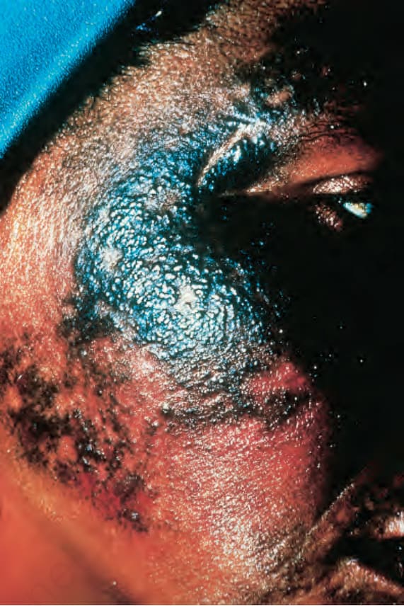
Fig. 13.136 Exogenous ochronosis: hyperpigmented plaque with numerous colloid milia in a Bantu female. The lesions developed as a consequence of the application of hydroquinone bleaching cream.
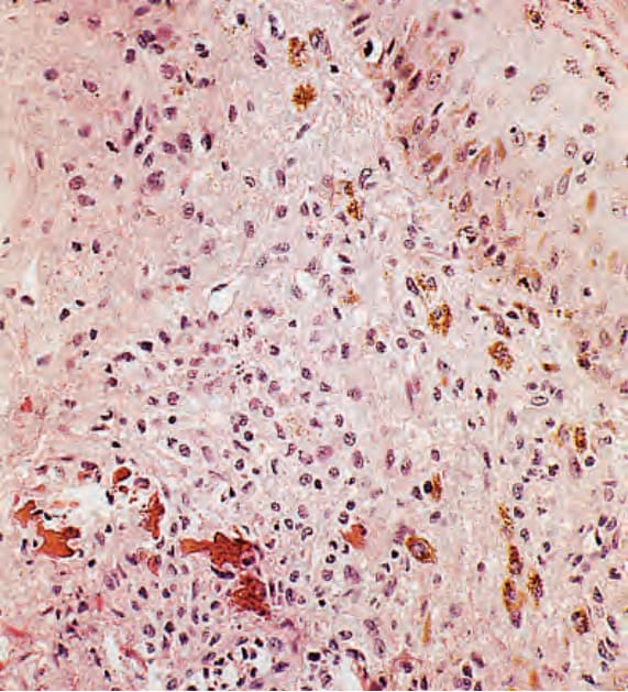
Fig. 13.137 Ochronosis: typical swollen, irregular, golden-brown fibers are seen (bottom left).
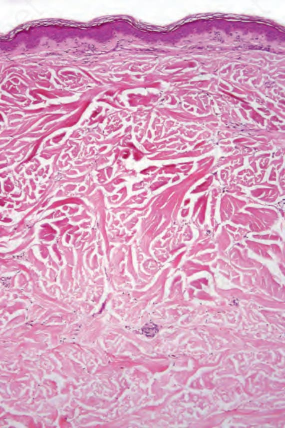
Fig. 13.138 Exogenous ochronosis: early lesion showing markedly swollen collagen fibers.