Hereditary coproporphyria
Hereditary coproporphyria
Clinical features This very rare autosomal dominant form of porphyria develops as a result of a deficiency of coproporphyrinogen oxidase.1,2 This enzyme catalyzes the sixth step in the heme biosynthetic pathway. It has been mapped to the long arm of chromosome 3 (3q11.2).3 A number of different mutations have
Increased hepatic iron stores are a major predisposing factor.6,20,31,32 The mechanism by which this happens is not well understood. Iron catalyzes the formation of reactive oxygen species and this may enhance uroporphyrin
594 Degenerative and metabolic diseases
formation by increasing the rate at which uroporphyrinogen is oxidized to uroporphyrin, leading to the manifestations of the disease. A second possible proposed mechanism considers the indirect inhibition of uroporphyrinogen decarboxylase by iron. Whatever the mechanism, the iron overload has important therapeutic implications as venesection can induce a remission.
Hepatitis C virus infection is often associated with porphyria cutanea tarda.32–35 A frequent association is also the acquired immunodeficiency syndrome (AIDS).36–43 AIDS patients with porphyria cutanea tarda are often hepatitis C virus-positive.43,44 Patients who have had both acquired and familial variants have developed the typical features of increased skin fragility, blistering, hyperpigmentation, and hypertrichosis, but scarring and milia have rarely been evident.45 Often, the development of porphyria has preceded the diagnosis of HIV infection.41 In many instances this has been related to excessive alcohol consumption and/or infectious hepatitis, particularly hepatitis C.40,46 The association has been reported too often to be merely fortuitous and liver damage seems to be the common denominator. The causal agent (be it hepatitis C virus or HIV) seems to have a direct effect upon hepatocyte porphyrin metabolism. It has been demonstrated that elevated serum porphyrin levels occur in early-stage HIV infection and hepatitis C infection.47 Porphyria cutanea tarda has also been described in association with nonalcoholic liver disease, chronic hemodialysis, noninsulin dependent diabetes mellitus and lupus erythematosus.1,48,49 An autoantibody study in a large series of patients with lupus erythematosus suggests that the association is fortuitous.50 The association with hematological malignancies, including leukemia and lymphoma, is usually related to the treatment, particularly repeated blood transfusions.51,52
Typically, blisters occur on light-exposed skin and are traumatic or actinically induced (Figs 13.101–13.103).4 Cutaneous fragility is usually marked. The blisters are slow to heal and leave superficial atrophic scars with milia. Although they are most often seen on the backs of the hands, they may also be found on the palms, face, scalp, forearms, trunk, and under the finger nails.4 Hypertrichosis and premature aging with chronic actinic damage are usual and sclerodermatous changes may be marked (Fig. 13.104). The hypertrichosis is characterized by long dark lanugo hair developing about the cheeks and temples, the eyebrows, ears, and arms (Fig. 13.105).4 The sclerodermatous features, which are more common in females, are found on both light-exposed and unexposed skin. Sites that are particularly affected include the face, neck, scalp, chest, and backs of hands, and often there is hyper- or hypopigmentation or both.4,53–55 In rare cases, patients may clinically present as scleroderma without other manifestations of porphyria cutanea tarda.55 Hyperpigmentation, if present, may be diffuse, reticulate or spotty. Preauricular calcification is a common complication. The dermal fibrosis appears to be related particularly to high uroporphyrin
levels.53 Uroporphyrin has been shown to stimulate fibroblast collagen synthesis independent of ultraviolet light.56
Uncommon cutaneous manifestations of porphyria cutanea tarda include alopecia affecting the frontoparietal, temporal, and occipital regions of the scalp, and centrofacial papular lymphangiectasis.57–59 Hair darkening has also been reported.60 Very rare cases have been documented presenting with plaques or simulating solar urticaria.61,62
Acute attacks are not a feature of this variant. Biochemical evidence of liver involvement is common, but clinical manifestations are unusual.1 Urinary porphyrin levels are increased and result in pink–red fluorescence with a Wood lamp.63
The diagnosis is confirmed by the presence of uroporphyrin and heptacarboxylic porphyrins in urine and plasma and by the presence of isocoproporphyrin in feces.
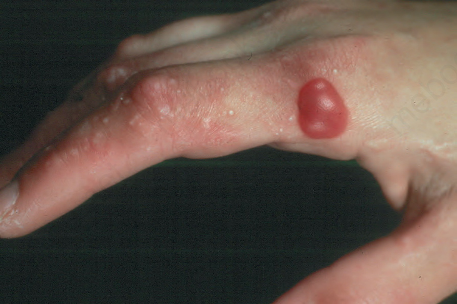
Fig. 13.101 Porphyria cutanea tarda: in addition to a blood-filled vesicle there are numerous milia. By courtesy of G. Murphy, MD, Beaumont Hospital, Dublin, Eire.
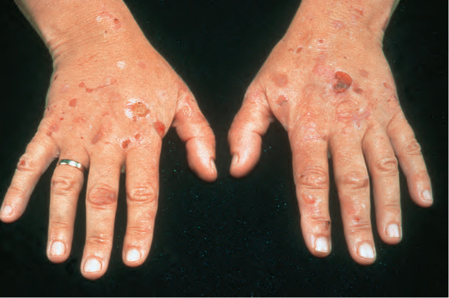
Fig. 13.102 Porphyria cutanea tarda: there are numerous ruptured blisters. Milia are also evident. By courtesy of the Institute of Dermatology, London, UK.
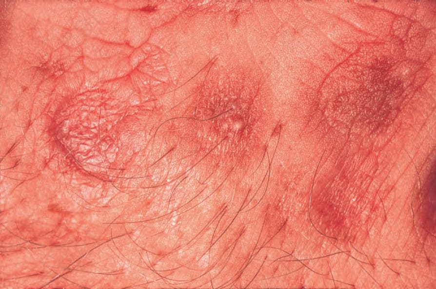
Fig. 13.103 Porphyria cutanea tarda: note the scarring and milia. By courtesy of the Institute of Dermatology, London, UK.
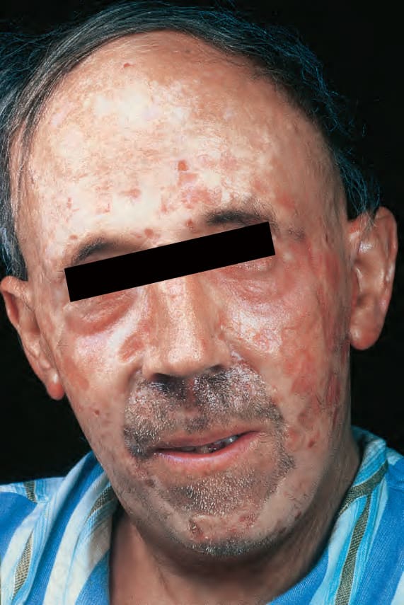
Fig. 13.104 Porphyria cutanea tarda: there is marked facial scarring with sclerodermiform features. By courtesy of G. Murphy, MD, Beaumont Hospital, Dublin, Eire.
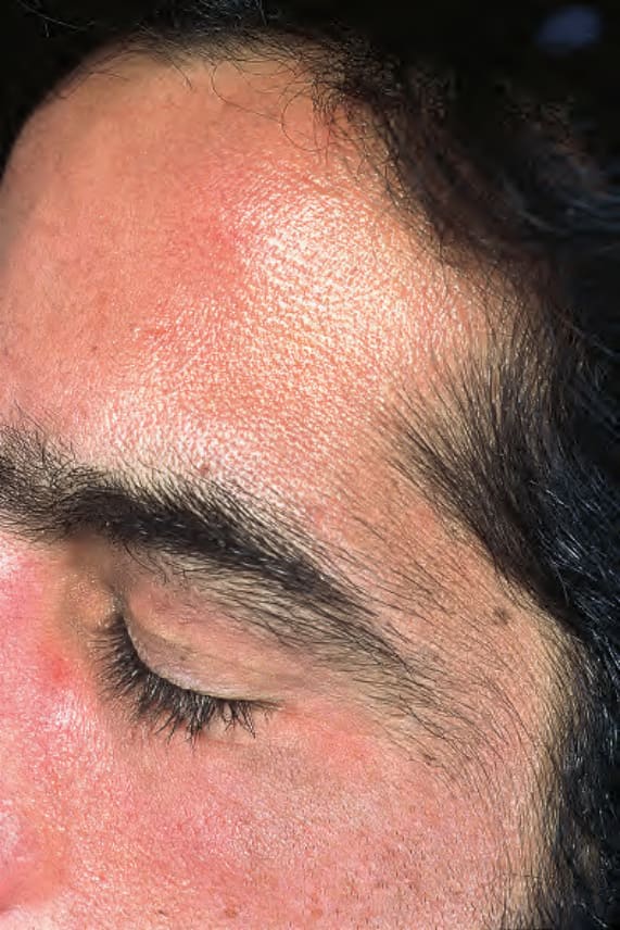
Fig. 13.105 Porphyria cutanea tarda: hypertrichosis as seen in this patient is a very typical feature. By courtesy of the Institute of Dermatology, London, UK.