Erythropoietic protoporphyria
Erythropoietic protoporphyria
Clinical features Although this condition was not recognized until 1961, it is now known to be the second commonest type of porphyria. It results from increased production of protoporphyrin due to diminished ferrochelatase (heme synthase) activity.1–3 Ferrochelatase is the enzyme responsible for the combination between protoporphyrin IX and iron to form heme. Urinary porphyrins are normal because protoporphyrins are insoluble in water. Protoporphyrin is elevated in plasma, erythrocytes, and occasionally in the feces.1 Coproporphyrins may be found in erythrocytes and feces. The mode of inheritance is predominantly autosomal dominant with incomplete penetrance although an autosomal recessive inheritance has also rarely been reported.4–6 The gene for ferrochelatase has been mapped to the long arm of chromosome 18 (18q21.3).7 A less common genetic variant is the X-linked dominant form
592 Degenerative and metabolic diseases
caused by a gain of function mutation in erythroid-specific 5-aminolevulinate synthase on the X-chromosome.8–12 This form appears to be more common in North Africa.11
The variable clinical manifestations of this disease are probably the result of heterogeneity of the ferrochelatase gene defects.4,13,14 Acute photosensitivity usually presents in early childhood.15 A painful burning erythema with edema occurs immediately after exposure to sunlight.16 Petechiae can occur, particularly with prolonged exposure. Vesicles are uncommon, but a scaly, erythematous reaction may be seen, leading to circular or linear depressed scars on the face (particularly on the bridge of the nose and around the mouth) and over the knuckles (Figs 13.95–13.99).1 Purpura and urticaria are sometimes seen. There may also be a waxlike thickening of the skin,
particularly of the dorsum of the hands and, more rarely, the face (Fig. 13.100).1 Bullae and milia have been documented exceptionally.3 A further case presented with pseudoainhum.17 An association with lupus erythematosus is very rare.18 Hypertrichosis and hyperpigmentation are not typically seen.2
593 Porphyria
been documented.4,5 Heterozygous patients often do not manifest symptoms of the disease.6 In those who develop symptoms, these usually appear after puberty. Affected patients develop intermittent attacks of abdominal pain in association with neurological and psychiatric manifestations. About 30% of cases develop photosensitivity, usually at the time of the acute attacks. The cutaneous changes are similar to those described for porphyria cutanea tarda. The disease may be precipitated by pregnancy, the contraceptive pill, fasting, infections, and the anabolic steroid methandrostenolone.3,7–9 Diagnosis is confirmed by the presence of increased excretion of coproporphyrinogen III in urine and feces. Porphobilinogen and aminolevulinic acid are increased during the episodic attacks.
Harderoporphyria is regarded as a variant form of hereditary coproporphyria in which hematological alterations predominate.2,10 Patients present with jaundice, severe chronic hemolytic anemia starting in the neonatal period, hepatosplenomegaly, and photosensitivity. Neuropsychiatric symptoms or abdominal pain are not seen. These patients usually have a specific mutation (K404E) on one or both alleles of the coproporphyrinogen gene.11
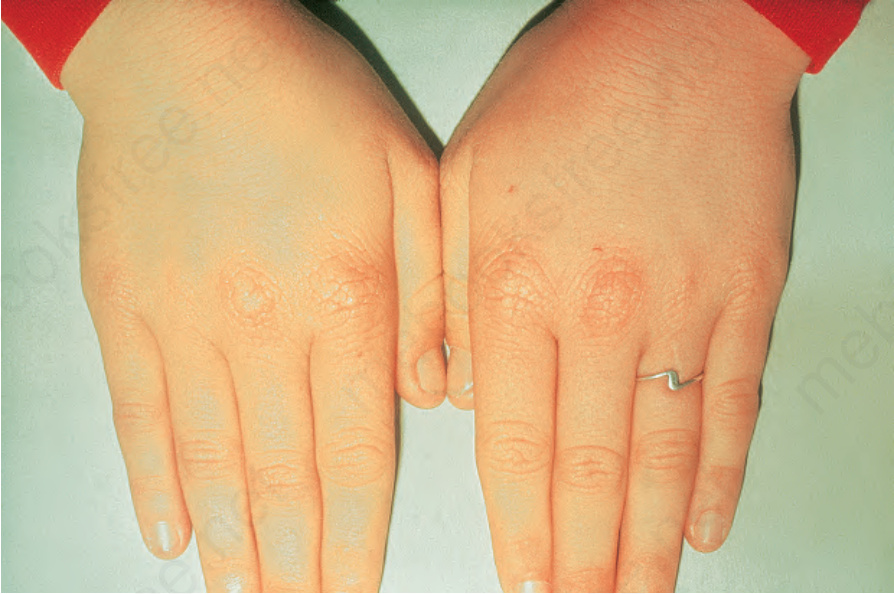
Fig. 13.100 Erythropoietic protoporphyria: there is characteristic waxy thickening of the skin of the hands. By courtesy of G. Murphy, MD, Beaumont Hospital, Dublin, Eire.
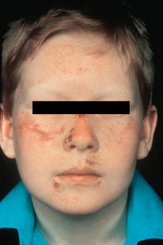
Fig. 13.95 Erythropoietic protoporphyria: crusted lesions are present on the cheeks, nose, and around the mouth. By courtesy of G. Murphy, MD, Beaumont Hospital, Dublin, Eire.
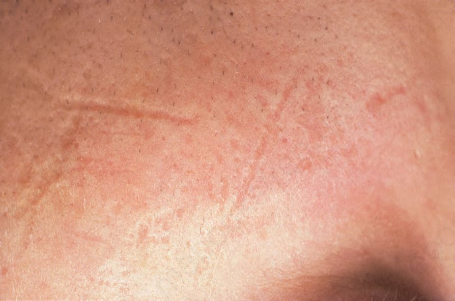
Fig. 13.96 Erythropoietic protoporphyria: there is marked scarring. Note the depressed linear lesions. By courtesy of G. Murphy, MD, Beaumont Hospital, Dublin, Eire.
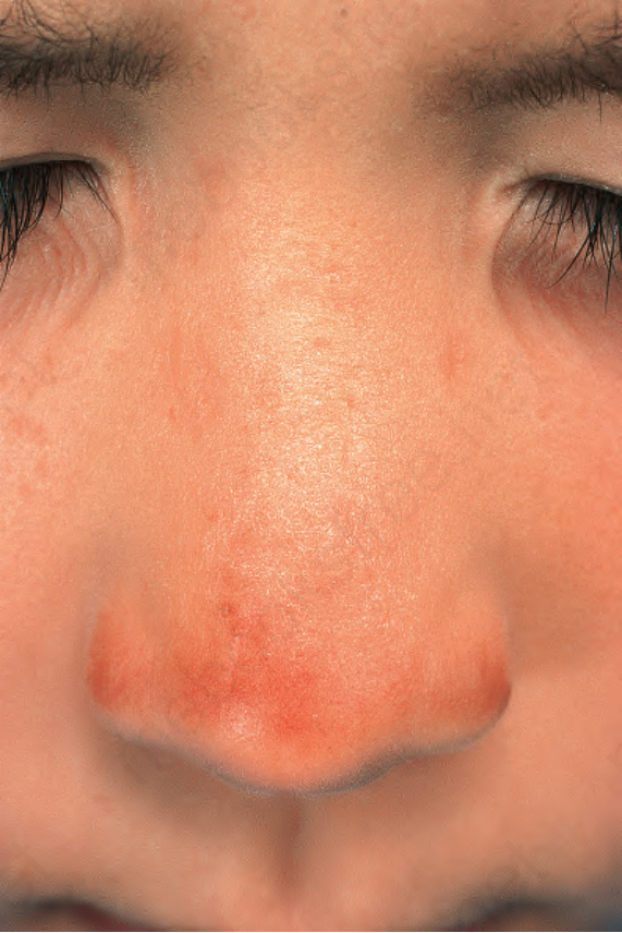
Fig. 13.97 Erythropoietic protoporphyria: there are characteristic, depressed, small linear scars on the bridge and sides of this patient’s nose. By courtesy of the Institute of Dermatology, London, UK.
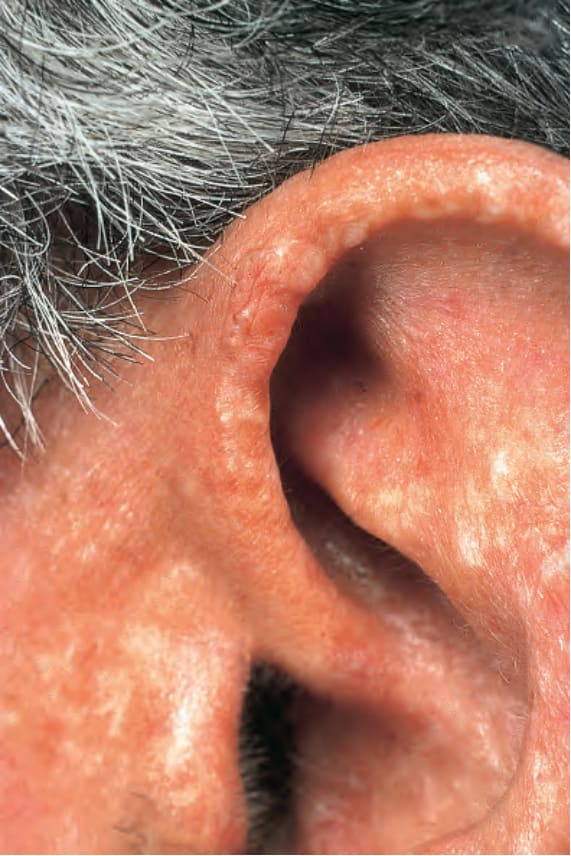
Fig. 13.98 Erythropoietic protoporphyria: there is very severe actinic damage. By courtesy of the Institute of Dermatology, London, UK.
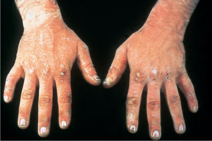
Fig. 13.99 Erythropoietic protoporphyria: note the characteristic scaly scars over the knuckles. By courtesy of the Institute of Dermatology, London, UK.