Cutaneous macroglobulinosis
Cutaneous macroglobulinosis
Clinical features Cutaneous macroglobulinosis (IgM storage papules) is a rarely documented manifestation of Waldenström macroglobulinemia.1–16 The latter is a chronic lymphoproliferative condition that typically presents in the fifth and sixth decades and shows a slight predilection for males.5 It is characterized by proliferation of lymphoplasmacytoid cells in the bone marrow, lymph node, and spleen and IgM paraproteinemia.5 Patients present with weakness, fatigue, weight loss, anemia, mucous membrane bleeding, retinal hemorrhages, lymphadenopathy, hepatosplenomegaly, peripheral neuropathy, and the hyperviscosity syndrome.7,8 Skin involvement is very uncommon and includes papules, nodules, tumors, plaques, and macroglobulinosis cutis. Additional features that are sometimes encountered include purpura, xanthomata, cryoglobulinemia, and Raynaud phenomenon.7
Clinically, macroglobulinosis presents as sometimes pruritic, skin-colored, erythematous or translucent papules measuring up to 1.0 cm in diameter distributed predominantly on extensor sites including knees, elbows, buttocks, and the arms and legs.7 Umbilication, erosion, and crusting and hyperkeratosis are commonly seen.5,9,10,11,15 Cutaneous tumor deposits present as violaceous nodules and plaques.
Histologic features The papules are characterized by homogeneous eosinophilic material in the papillary and reticular dermis (Fig. 13.89).2,11 Hair follicles and eccrine glands may be encased.2,11 The deposits are PAS positive but are Congo red negative (Fig. 13.90). Vessels may also show occlusion by the same
589 Porphyria
material.11 A lymphoplasmacytoid infiltrate is variably present.7 The plasma cells may contain intracytoplasmic IgM-rich vacuoles.4
Direct immunofluorescence and immunohistochemistry show that the deposits stain strongly for IgM.2,5,7,11
Ultrastructurally, the deposits are composed of amorphous or granular and sometimes filamentous material which by immunoelectron microscopy consists of IgM.1–3,7 The periodicity of amyloid is absent in the filamentous component.7
The plaques and tumor nodules are composed of lymphoplasmacytoid infiltrates.
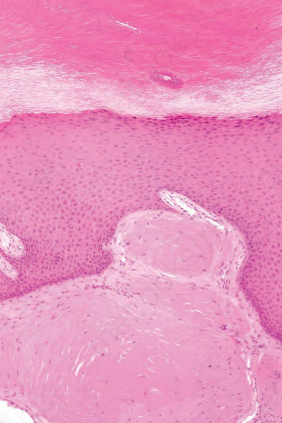
Fig. 13.89 Macroglobulinosis cutis: these are nodular deposits of eosinophilic material in the superficial dermis. By courtesy of A. Wang, MD, Brigham and Women’s Hospital, Boston, USA.
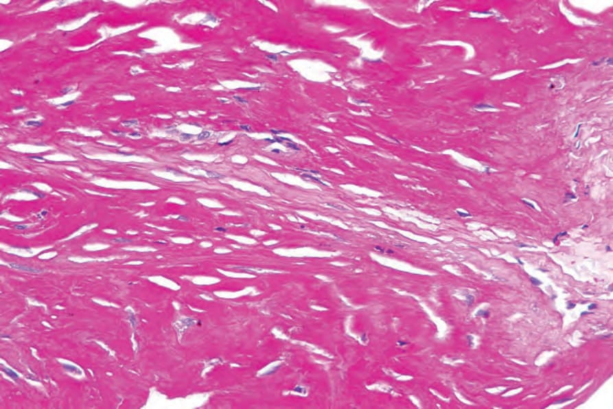
Fig. 13.90 Macroglobulinosis cutis: the material is strongly periodic acid–Schiff positive. By courtesy of A. Wang, MD, Brigham and Women’s Hospital, Boston, USA.