Adult colloid milium
Adult colloid milium
Clinical features This variant, which is much commoner than the childhood form, affects middle-aged patients and shows a predilection for males. Outdoor workers are most often affected and lesions seen on sun-exposed skin are often accompanied by the features of solar elastosis, giving rise to the synonym of papular elastosis.
Adult colloid milium presents as dome-shaped yellowish translucent papules measuring 0.1–0.5 cm in diameter and, in common with juvenile colloid milium, they contain gelatinous material. Lesions are most often seen on the face, ears, neck, and the dorsum of the hands (Fig. 13.78) and
Pathogenesis and histologic features In contrast to the keratinocyte changes seen in the juvenile variant, adult colloid milium represents an extreme degree of actinic damage centered upon the upper dermal elastic fibers. Although earlier studies suggested that the colloid might have represented abnormal collagen or a fibroblast secretory product, more recent studies suggest that it derives from actinic elastoid.17,23–25
Ultrastructural studies have shown that there is direct continuity between actinic elastoid and the colloid deposits and that, within the electron-dense
585 Hyalinosis cutis et mucosae
label with antikeratin antibodies, and immunoglobulins and complement are absent.
Histologically, the eosinophilic amorphous, autofluorescent clefted deposits are typically separated from the epidermis by a grenz zone containing normal collagen (Figs 13.79 and 13.80).24 Fibroblasts often occupy the fissures between the fragmented deposits.24
colloid, remnants of both normal and elastotic fibers may sometimes be identified.24,26 Amyloid filaments are not present. Further support for this hypothesis is given by the identification of SAP component within the colloid deposits.24 Although this protein is characteristically present within amyloid, it is also a constant component of normal elastic tissue and has also been identified in actinic elastoid.27,28 Adult colloid milium does not
Histochemically, adult colloid milium is diastase-resistant, PAS positive, thioflavine-T positive, and demonstrates apple-green birefringence with Congo red.17 It is also Dylon positive.2 Colloid milium can be difficult to distinguish from amyloidosis, and electron microscopy may be necessary.29,30
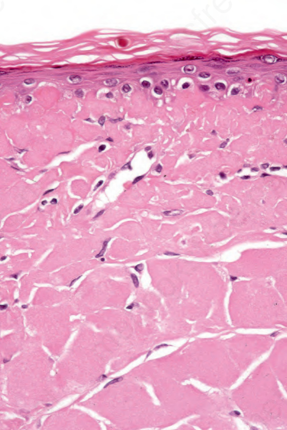
Fig. 13.75 Juvenile colloid milium: this high-power view shows the faceted nature of the deposit.
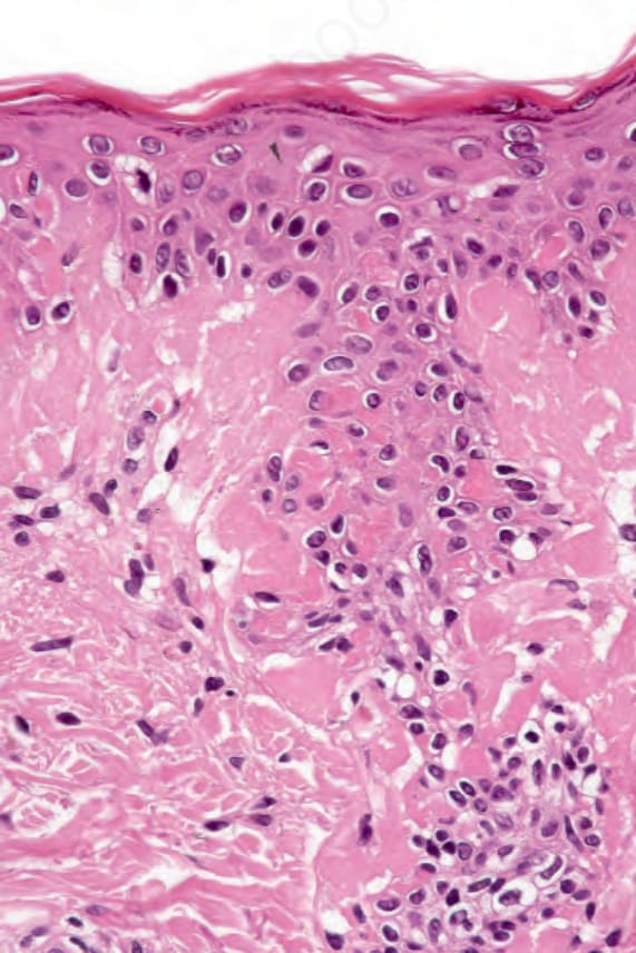
Fig. 13.76 Juvenile colloid milium: the adjacent epidermis shows massive apoptosis.
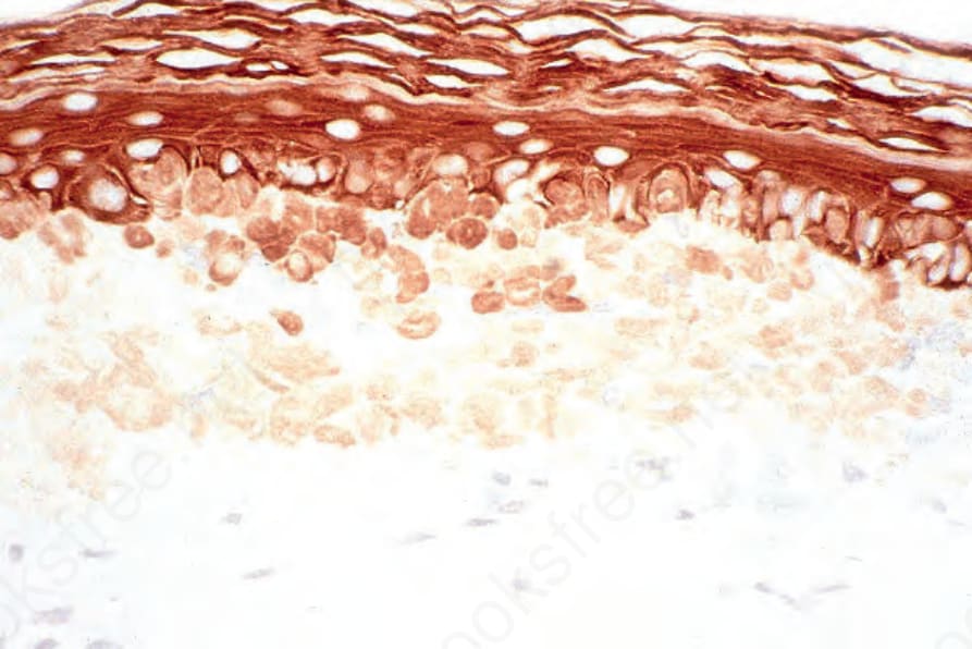
Fig. 13.77 Juvenile colloid milium: the amorphous material that characterizes this condition is of epidermal derivation. Tonofilaments undergo filamentous degeneration (apoptosis). Note the keratin positivity of the colloid aggregates (pankeratin).
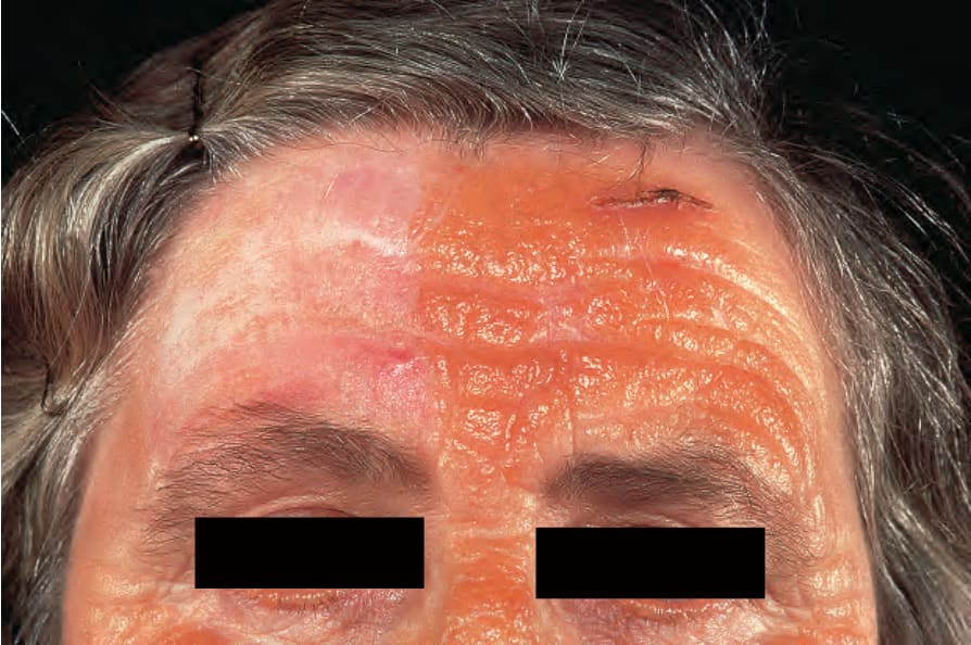
Fig. 13.78 Adult colloid milium: predominantly unilateral, streaked, orange plaque involving the forehead and nose. By courtesy of the Institute of Dermatology, London, UK.
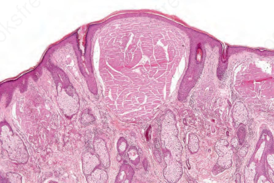
Fig. 13.79 Adult colloid milium: deposits of eosinophilic material are present in the superficial dermis. There is adjacent solar elastosis.